WARNING: GRAPHIC IMAGES ON THIS PAGE
⚠️
WARNING: GRAPHIC IMAGES ON THIS PAGE ⚠️
Read about the interesting cases that have come through our doors
Sonic's New Smile
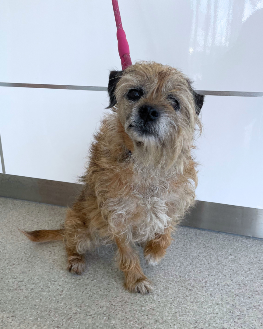
Sonic is a 10-year-old Border Terrier who had an ulcerated lump on the right side of her muzzle. As the lump was starting to grow with time, a Fine Needle Aspirate (FNA) test was recommended to determine the nature of the lump, and therefore plan for surgery if required.
The FNA results came back as a suspected Basal Cell Tumour, which is a typically benign lesion that is cured by complete surgical excision. Sometimes, however, they can exhibit malignant behaviour (i.e. invade deep into surrounding tissues), so removal was strongly recommended.
Due to the location on her muzzle, and the lack of extra tissue to aid in closure, we needed to utilise a skin flap. Skin flaps use “donor skin” from near the surgical site to close the “recipient site” with the blood supply intact.
In Sonic’s case, Dr Cindy assessed the lump and determined it would be possible to utilise a tension-free labial advancement flap, pulling the lip towards her nose to allow for closure.
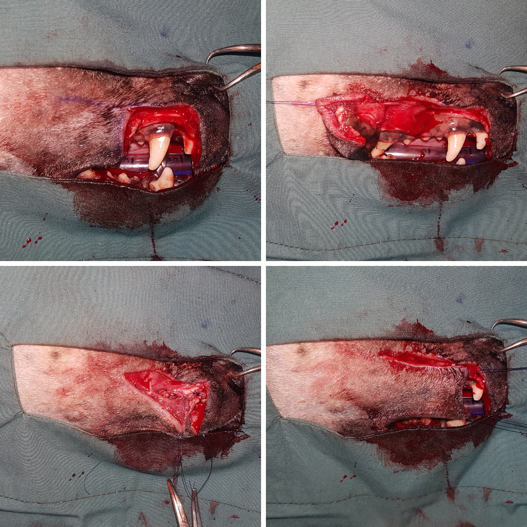
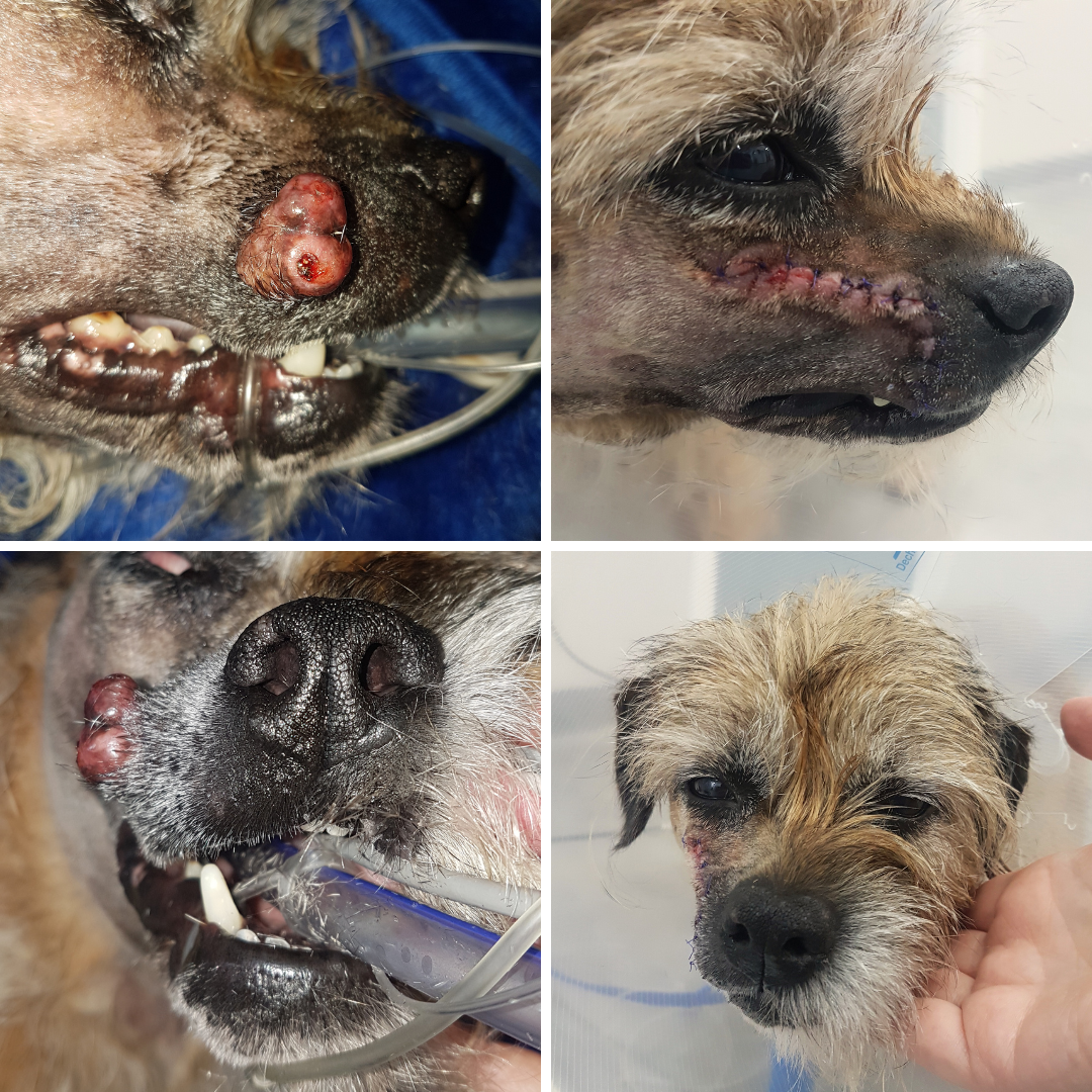
The surgery was successful, and the lump was sent to the lab for further testing. Despite the initial FNA result, the histopathology result came back as a trichoblastoma, which are benign tumours of the hair follicle, and are cured with surgical excision.
As Dr Cindy was able to attain appropriate margins, the trichoblastoma was fully removed, and therefore will not return in that location.
Sonic’s wound healed very nicely, and her scruffy fur eventually grew back!
Dinah's Rocking Bladder
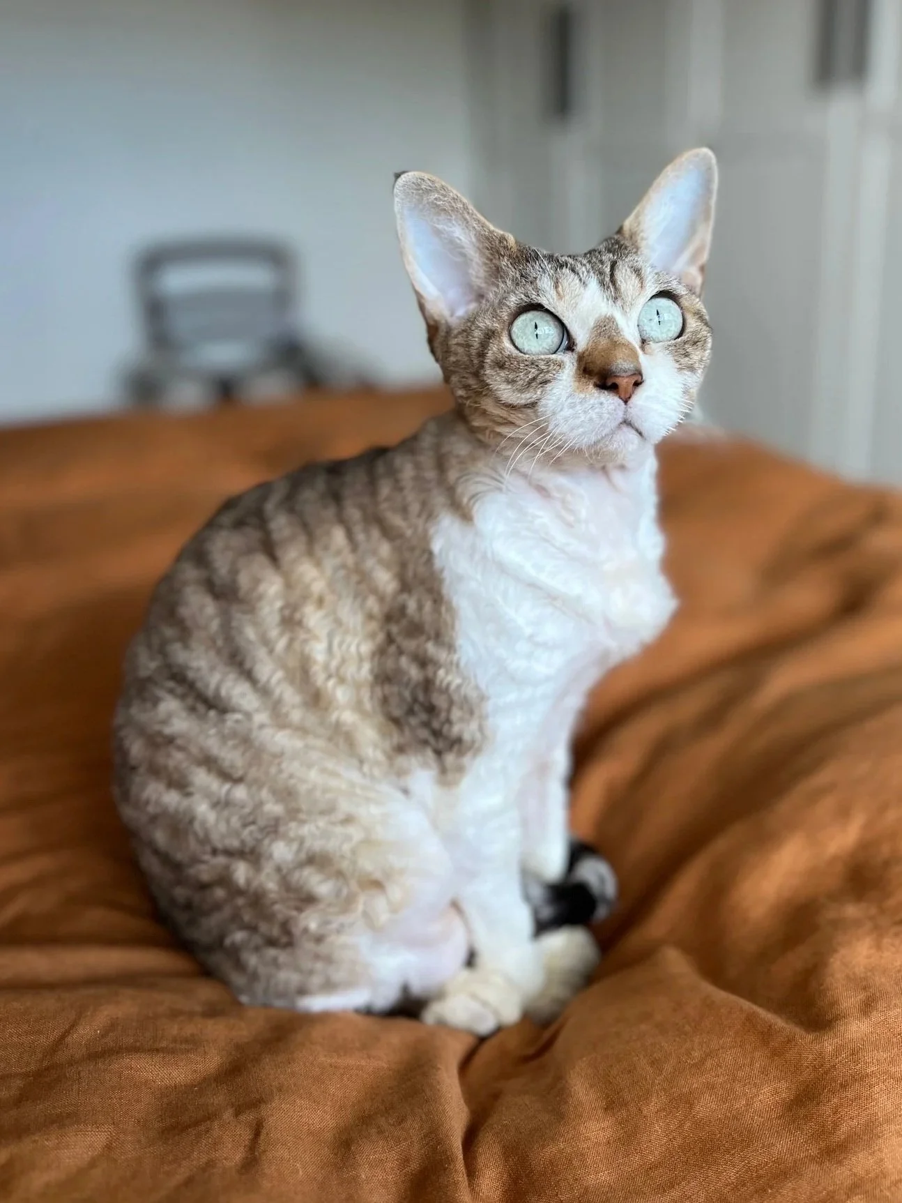
In 2022, Dinah had blood in her urine; suspected to be caused by feline interstitial cystitis (stress related bladder inflammation) due to the addition of a new puppy at home. The blood resolved after a course of oral anti-inflammatories (the treatment of choice for stress related cystitis).
A few months later her symptoms recurred, but this time medication did not help. Her urine sample was sent to the external lab for culture which showed no bacteria present but there were lots of blood and white blood cells (indicating inflammation).
Dinah had a specialist ultrasound of her bladder which showed she had two oval shaped uroliths (bladder stones) present.
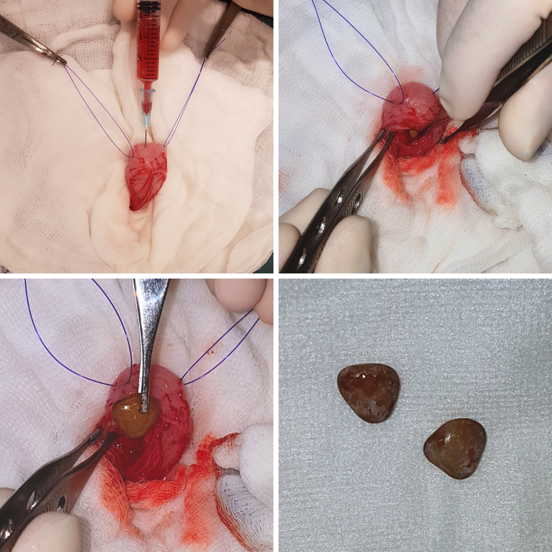
A cystotomy was performed to remove these stones. This surgical procedure involves an exploratory laparotomy (open abdominal surgery) to expose and open the bladder to retrieve the bladder stones. During the surgery Dr Cindy removed 2 triangular uroliths.
Dinah recovered quickly from surgery and was back to her normal self very quickly. She was commenced on Hills Prescription Diet k/d to assist in dilution of her urine and prevent her urine becoming too acidic.
Unfortunately (as is very common with patients with bladder stones) in June 2023 Dinah presented with blood in her urine again. Dinah was referred to the Animalius specialist medicine team to customise a management plan to prevent recurrence of bladder stones.
In August 2023 she underwent another cystotomy with Dr Jess who removed a large smooth bladder stone and Dinah then commenced her plan created by the Animalius team (medications to increase the pH of her urine and additional water added to her food to dilute her urine).
Squid's Eyeless Journey
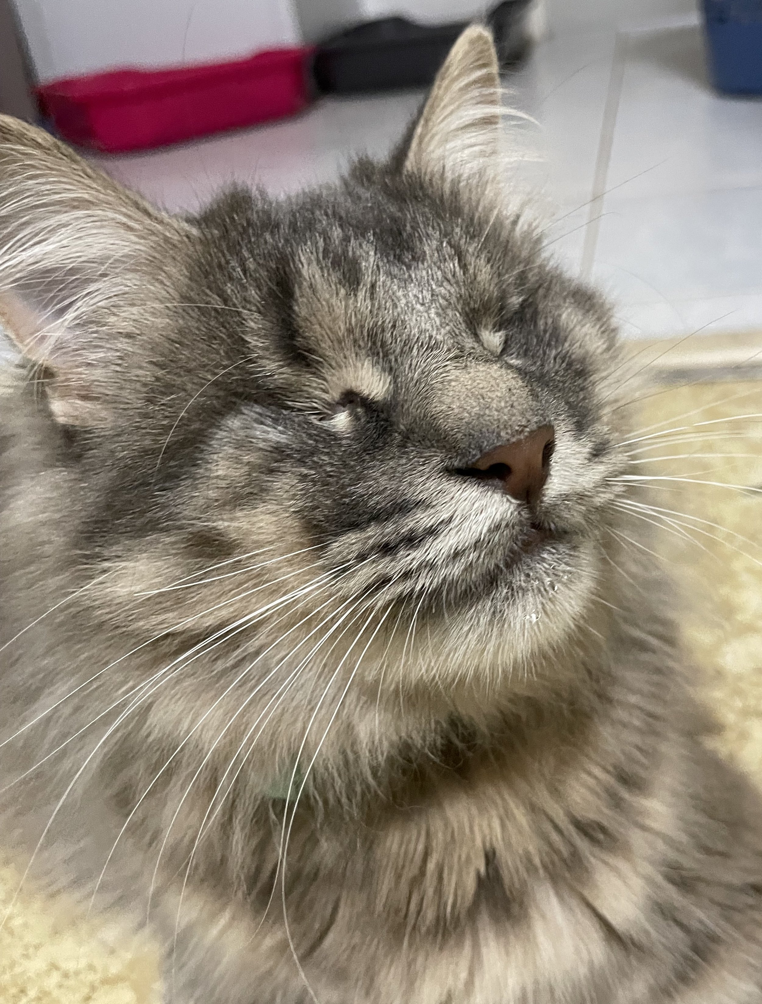
We first met Squid when he was just around 10 weeks old as a stray who was brought into KSAH. Squid was quite skinny at the time, with a belly full of worms and had very crusty and infected eyes. Upon further inspection, it became apparent that Squid had a condition known as “eyelid agenesis”.
Eyelid agenesis is a birth defect where a portion of the eyelid margin is underdeveloped. In Squid’s case, he was missing the outer half of both of his upper eyelids. Animals with eyelid agenesis usually are unable to blink correctly, and often have chronically sore eyes (due to hair rubbing on the eyes) and further eye abnormalities that could lead to blindness.
We suspected that Squid had very minimal to no vision in his eyes due to the way he interacted with his surroundings, and they were going to be a source of discomfort for him in the long term.
Larissa, our Customer Relations Officer with a preference for vision-challenged cats, fell in love with Squid and decided to adopt him.
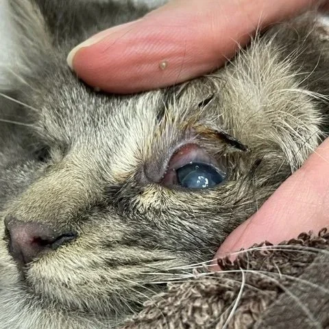
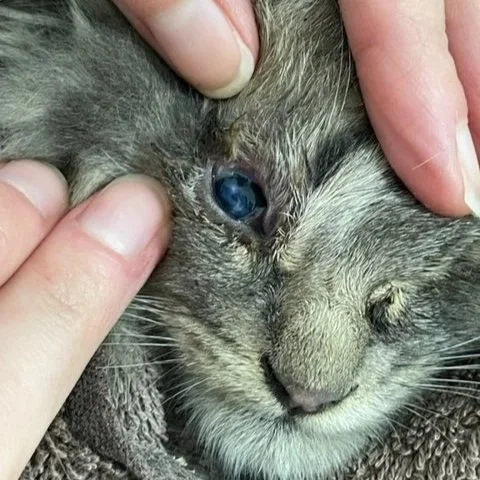
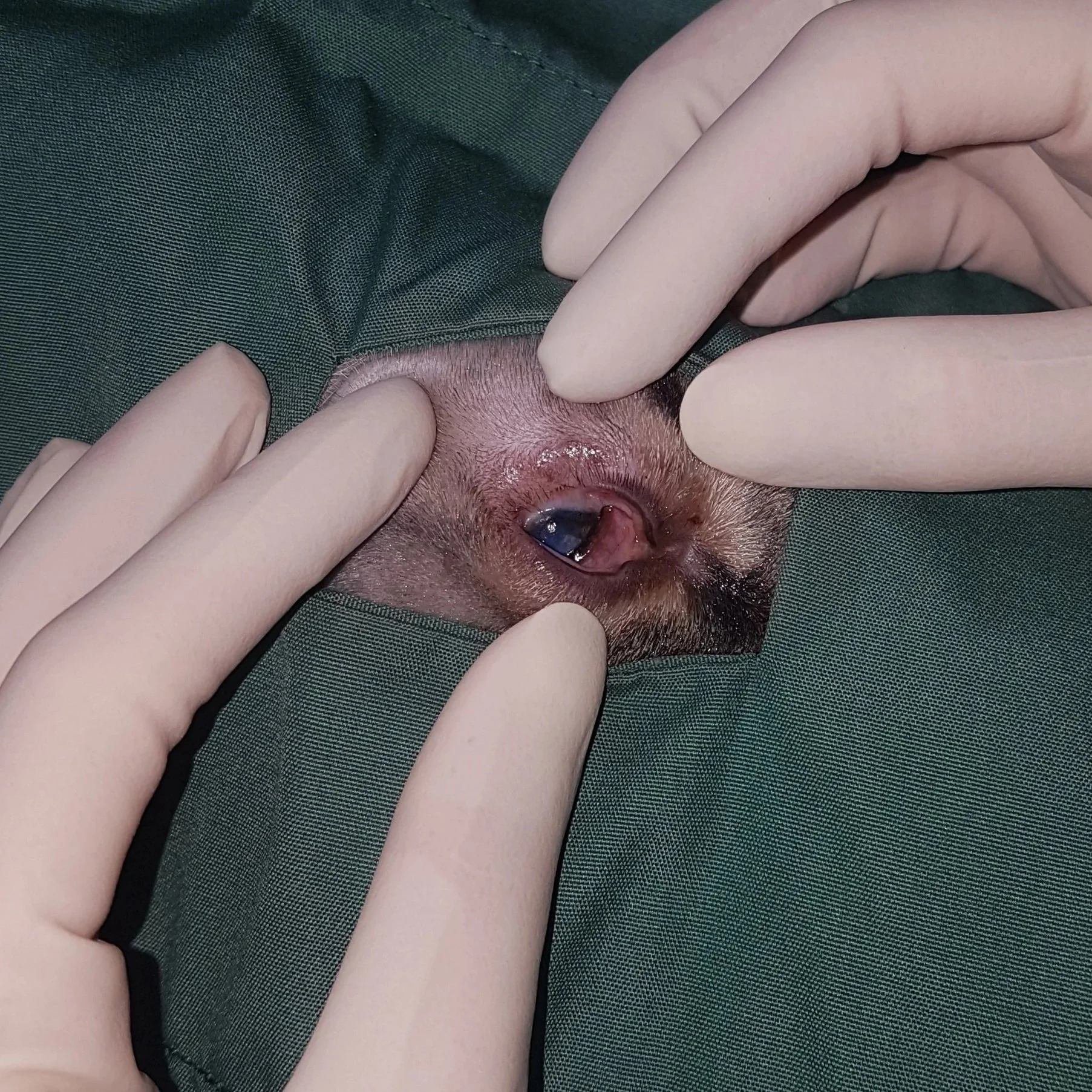
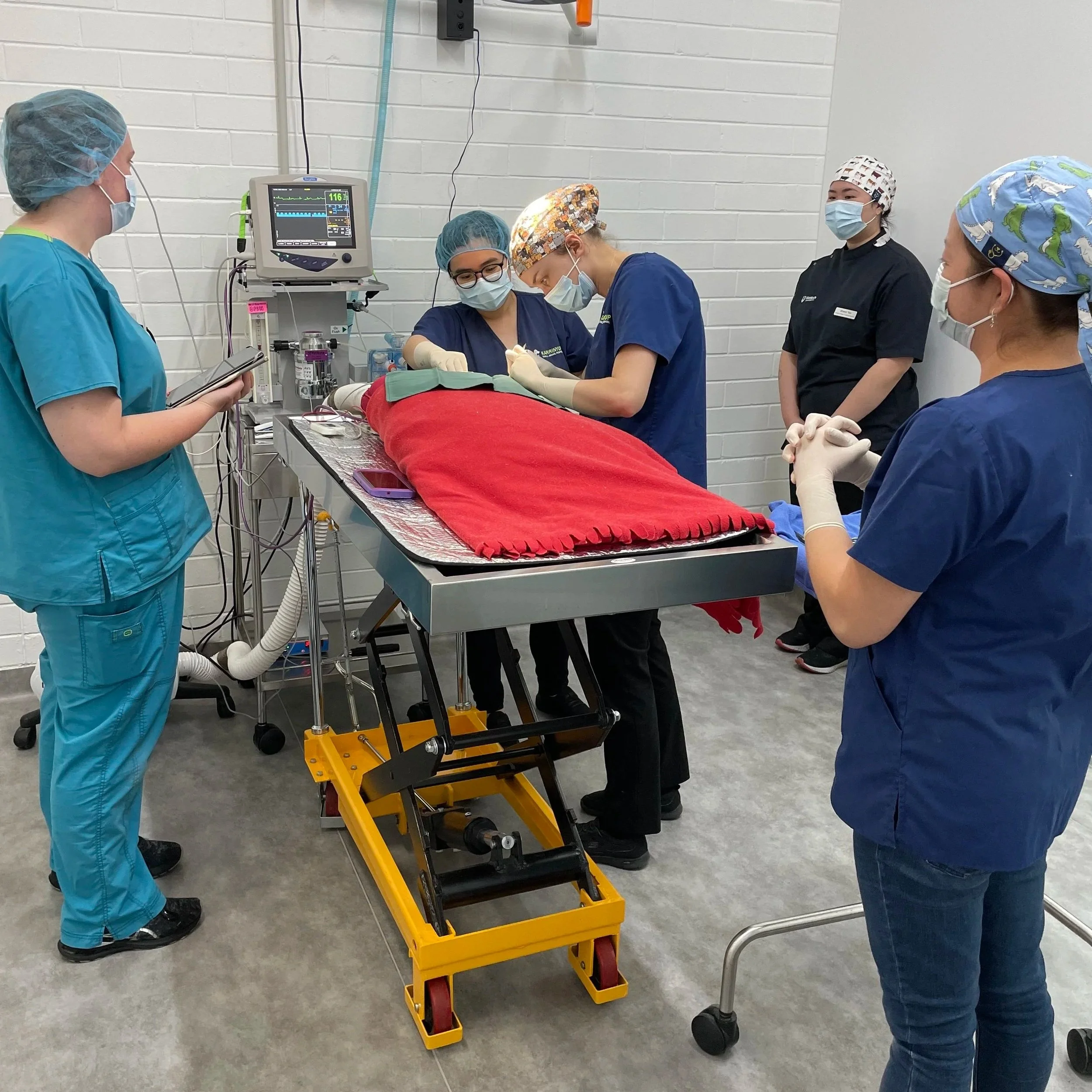
Medicating Squid became a challange, he was a semi-feral kitten, that needed eye medication multiple times a day. Once he was old enough to tolerate a long general anaesthetic, he was booked in to undergo a bilateral eye enucleation (removal of both his eyes). Dr Kristy and Dr Jess (under the very close supervision and guidance of Dr Cindy) together performed his surgery.
Since having the surgery, Squid is much more happy and comfortable living his day to day life, and enjoys the company of his blind sister Althy and cheeky brother Bort. Between the three of them they share 2 eyes!
Alfie's Tail Troubles
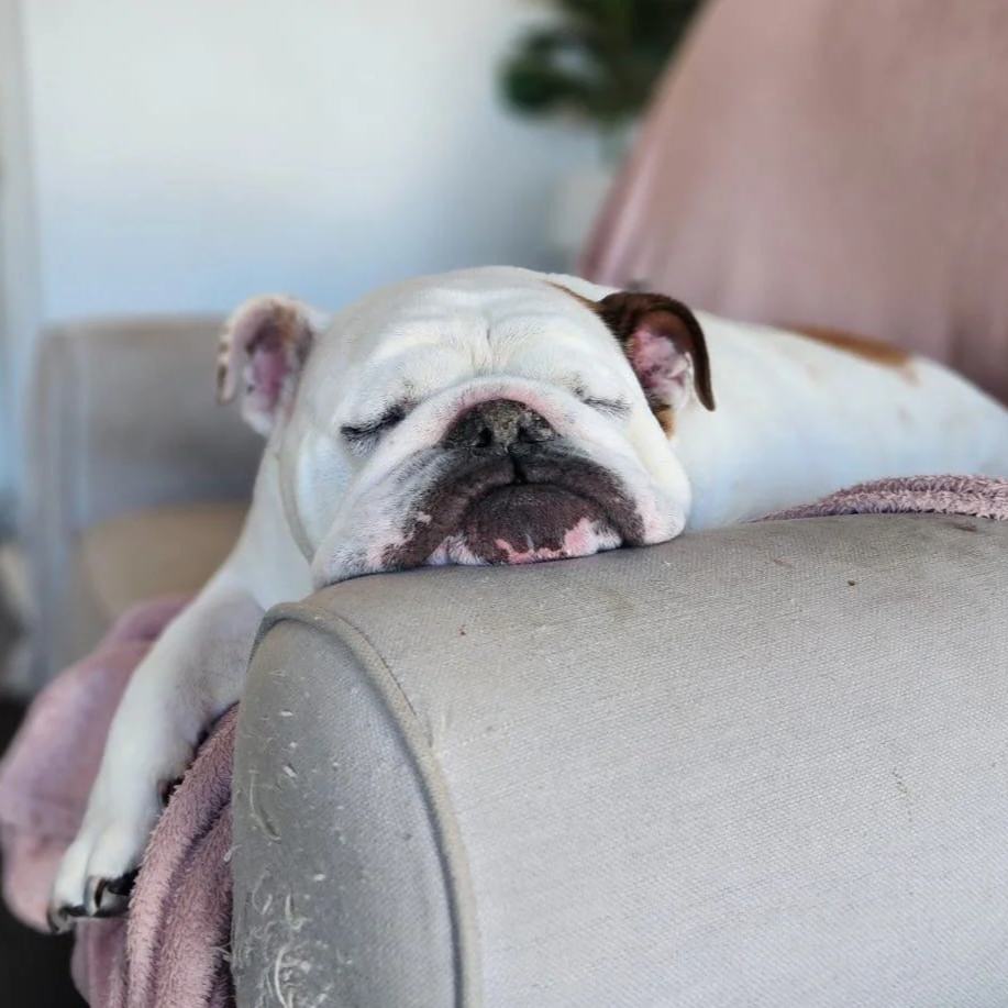
Alfie is an 8yr old bulldog who is a much loved patient of KSAH (and one of Dr Jess’s regulars).
Alfie has a long standing history of skin and food allergies which have resulted in secondary skin and ear infections in the past. His skin is relatively well managed at the moment with regular medicated washes to manage his secondary infections and cytopoint injections when he gets itchy.
Being a bulldog, his owners have needed to regularly clean all of his folds his entire life (including his facial folds and ears) but he began to resent the cleaning of his tail fold. His tail fold was caused by a combination of his skin rolls and his deeply recessed and very tight corkscrew tail which was pushing into the skin above his rectum. Due to the difficulty in keeping this deep fold clean, he began to develop painful skin infections and he became very sensitive around his back end.
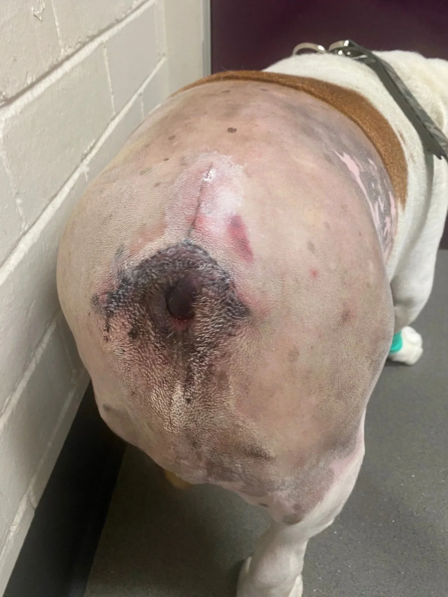
Dr Jess had discussions with Alfie’s owners about considering a tail amputation and resection of the skin fold due to how painful and bothered he had become. Due to the delicate and challenging nature of the surgery (as the tail base is very close to the rectum, blood vessels and nerves), Alfie was referred to the surgical department at Perth Veterinary Specialists.
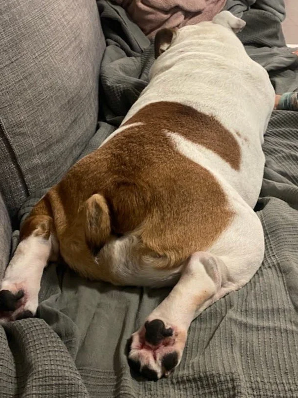
After assessment by their surgical team and a CT scan to determine the severity of his corkscrew tail, Alfie proceeded to have his deformed tail removed with Dr Tim Caporn. His surgical procedure went very smoothly and he recovered well in the weeks following surgery.
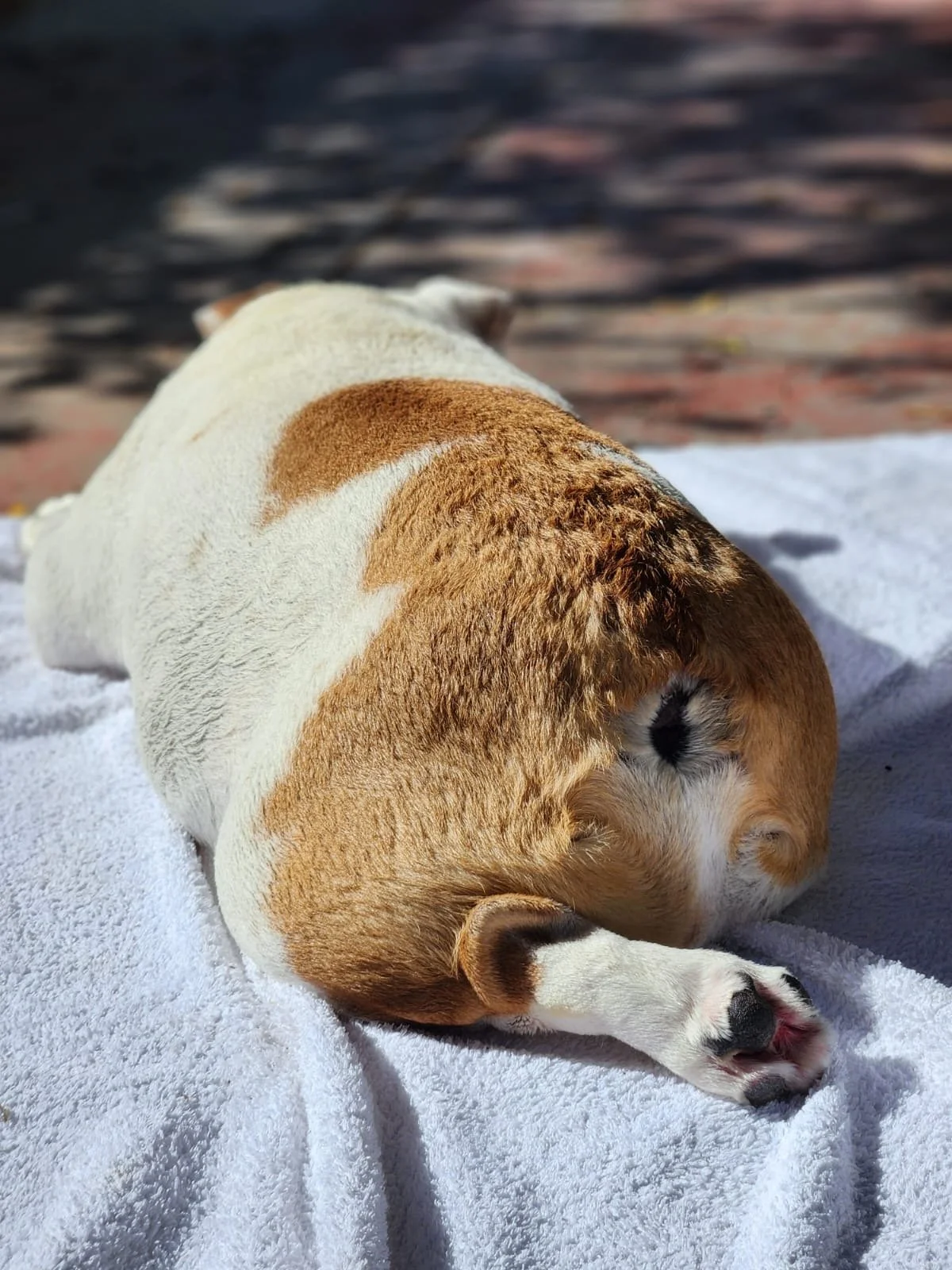
It has now been over 12 months since Aflie had his tail amputated and his owner reports that he couldn’t be happier. Since surgery he has been much brighter in himself and more energetic, indicating that his tail was likely causing him significant pain/discomfort.
Huan's Unfortunate Outing
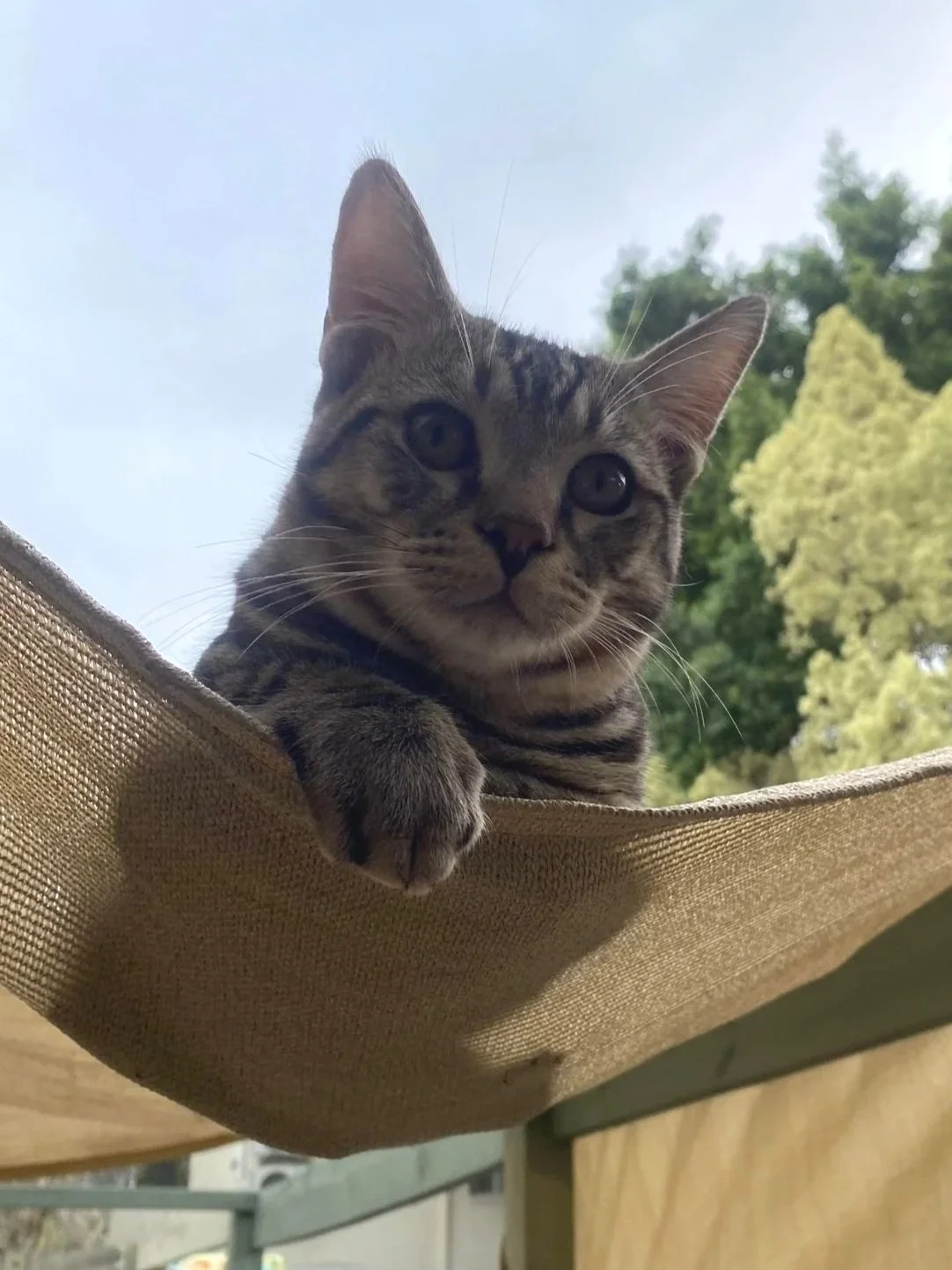
Huan is a 7 month old domestic shorthair cat who found himself on the wrong end of a mysterious accident. Huan’s mother found him bloodied and quiet, hiding under the bed and not wanting to eat. What had caused this however, was a mystery. Huan had suffered some trauma whilst playing outside, likely an accident with a passing car, but we will never know for sure.
On presentation to the hospital, Huan had numerous injuries throughout his body. Blood was found on all 4 paws, and his front left leg was sore and swollen. Fortunately, X Rays would later confirm that these injuries were superficial, with no fractures or damage to his vital structures, and no major injuries to his internal organs.
Huan’s face however, bore the brunt of the damage. One of his upper canine teeth had snapped in half, exposing the nerve and root structures, causing significant pain. This tooth had to be removed surgically to prevent infection and further complications. He had also sustained a deep laceration to his tongue which needed to be stitched back together.
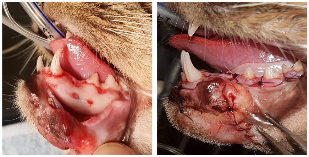
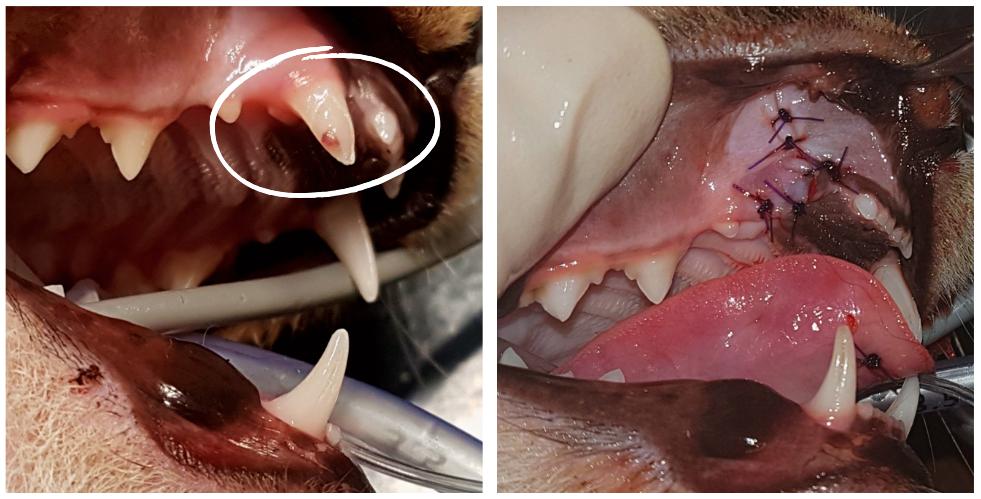
Most significantly however, the skin and outer gum layer had been completely torn away from one side of his lower jaw, exposing the underlying jaw bone. Such a sight is not for those with a weak stomach.
The major difficulty with surgically repairing such an injury, aside from the delicate nature of the surgery itself, is that the damage to the tissues themselves can cause them to die off leading to a breakdown of the surgical site. Fortunately for Huan, the gum and skin tissues remained healthy, and were successfully reattached to the underlying bone where they quickly healed.
Huan had to have a soft food diet for several weeks to allow his mouth to heal. In the end, he was lucky. Despite receiving some pretty horrific injuries, the poor little guy was fixed and was back to his normal self in quick time. Photos from just 15 days after the initial surgery show minimal evidence that the injury had ever taken place, and his mother reported that he was eating and drinking like nothing had happened. We can only hope he has learned some road safety after his ordeal.
Rosie's Ovariectomy
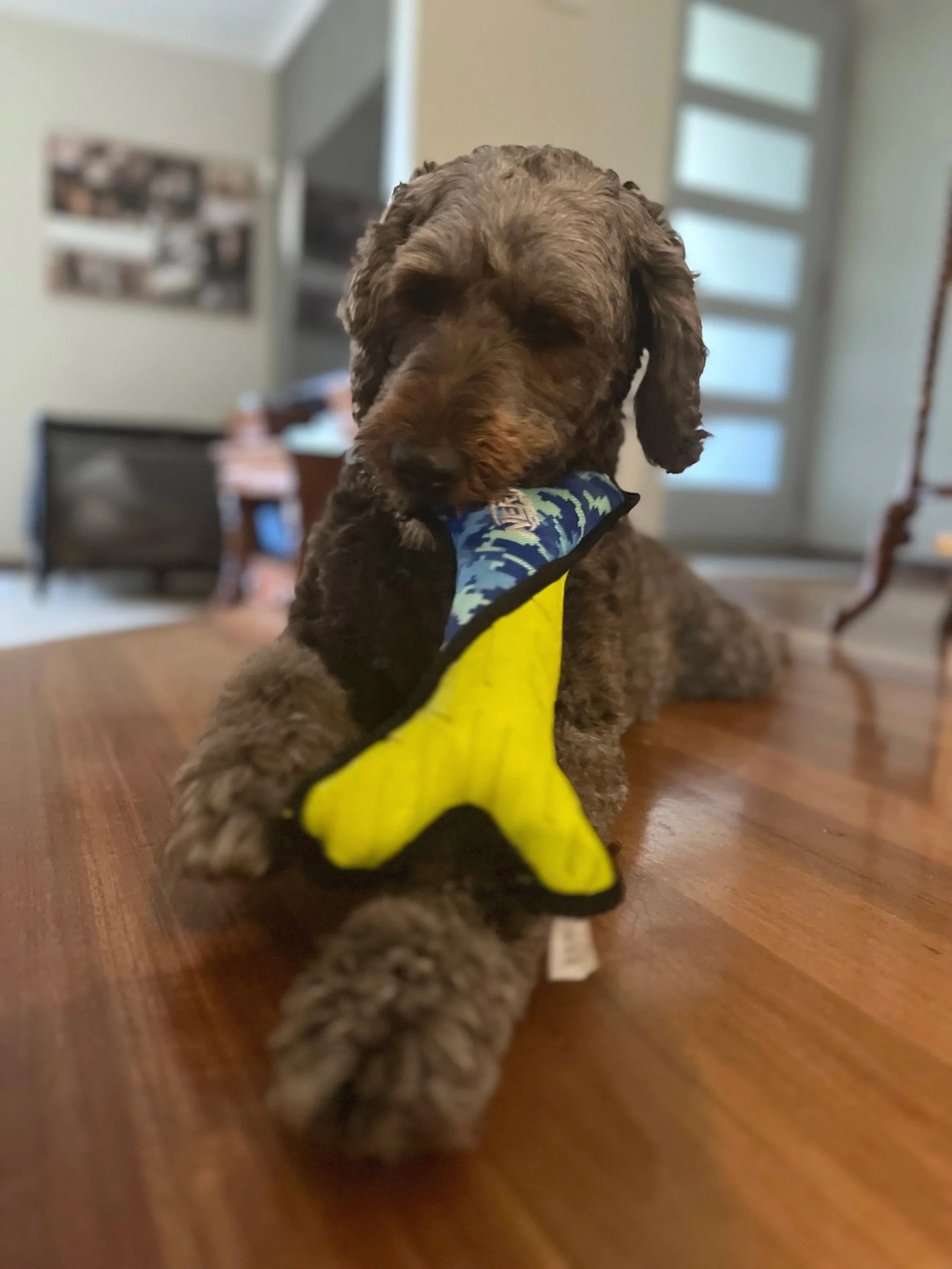
Rosie is a 4 year old Labradoodle who came to see Dr Cindy for abnormal vaginal discharge and frequent licking of her vulva.
Prior to coming to us, Rosie had an ovary sparing spay (a procedure involving removal of the majority of the uterus whilst leaving one or both ovaries intact). This procedure is occasionally performed instead of the usual ovariohysterectomy (removal of the uterus and both ovaries), as it allows for continued hormone production and regular cycling without the possibility of pregnancy. Dogs that undergo ovary sparing spays, however, can have an increased risk of ovarian and mammary cancers, uterine infections, and ongoing sexual behaviours, compared to dogs that have had ovariohysterectomies.
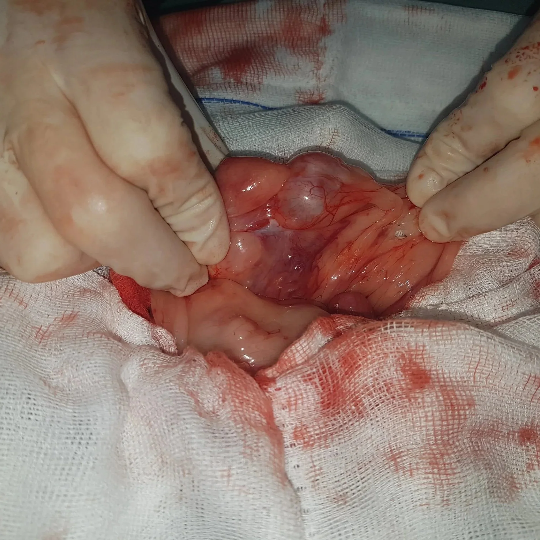
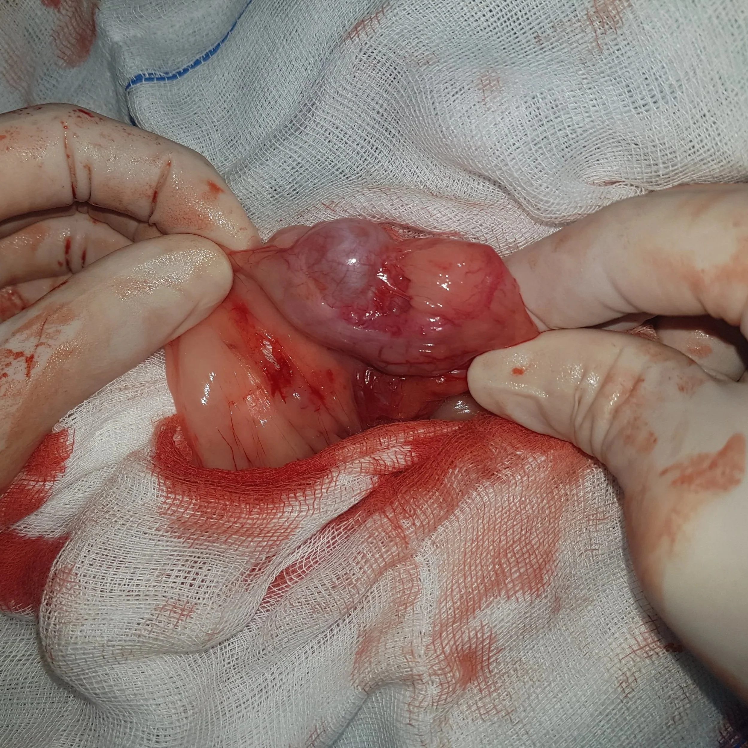
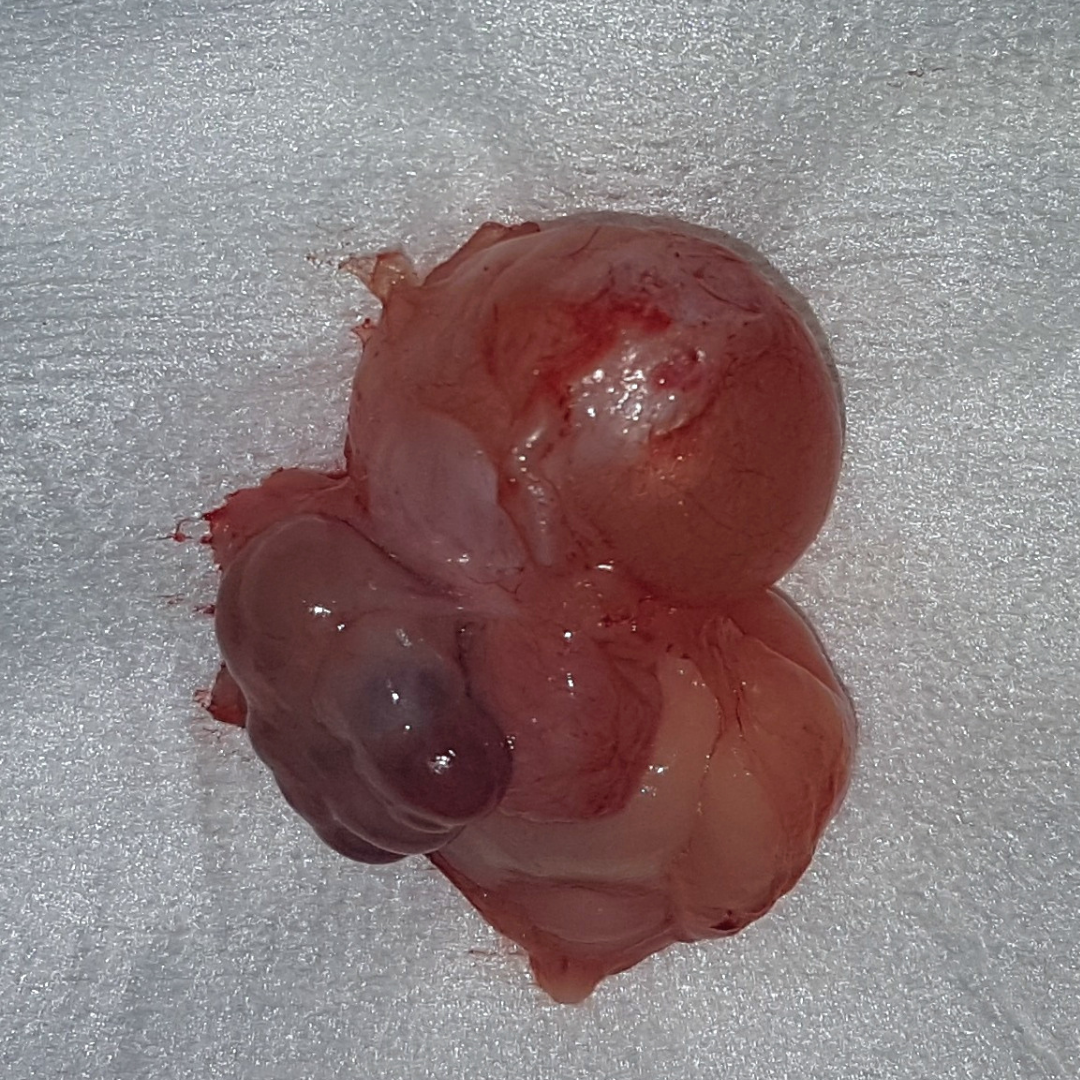
In Rosie’s case, it was suspected that her problems were due to either infection of the vagina or the remaining uterine stump, brought on by her continued hormone production. An abdominal ultrasound was performed to assess her remaining left ovary, uterine stump, and to look for any remnants of her right ovary. The ultrasound found that the left ovary had a number of large fluid-filled cysts, called a polycystic ovary.
Polycystic ovaries occur when the ovary is unable to ovulate the follicles it produces during a heat cycle. This results in signs of persistently being on heat, such as vulvar swelling, bloody or mucousy vaginal discharge, and behavioural changes. If the dog is then induced to ovulate with medication, there is a strong likelihood she will get a uterine or vaginal infection, so the preferred treatment is removal of the ovary. Dr Cindy removed Rosie’s left ovary, and sampled the vaginal discharge to determine if an infection was present.
Because she had been persistently on heat, there was significant bleeding during surgery (increased blood supply to the reproductive organs), and fibrous adhesions in the abdomen due to her previous surgery. We are equipped with an electrocautery system that allowed us to efficiently stop the bleeding, and the rest of the operation went well.
Results from Rosie’s vaginal swabs also showed a concurrent vaginal infection that was treated with a course of antibiotics.
Sophia And Her Nuclear Vacation
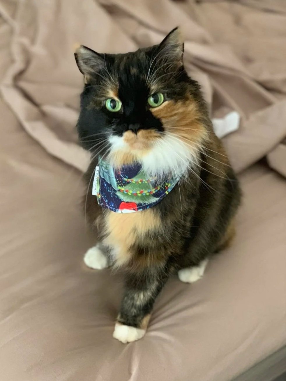
Sophia is a 15 year old domestic medium hair cat who belongs to Nurse Emma. Unfortunately, last year she was diagnosed with hyperthyroidism.
Hyperthyroidism is most common in older cats and can have a number of symptoms. Usually, it is caused by a benign growth in the thyroid known as an adenoma. This enlarged thyroid then produces excess thyroid hormones, resulting in a greatly increased metabolism and high blood pressure. In the early stages, we often see patients lose weight despite having an increased appetite. Patients can have an unkempt coat, have vomiting, diarrhoea, or become hyperactive. If left untreated, multiple organs (eg. heart, eyes) may become affected and deteriorate over time. Diagnosis can be confirmed with a blood test.
Once diagnosed, treatment can commence. Most commonly, long term twice daily medication is the treatment of choice for many patients. However, this option still carries the risk that the usually benign adenoma could develop into malignant cancer (adenocarcinoma).
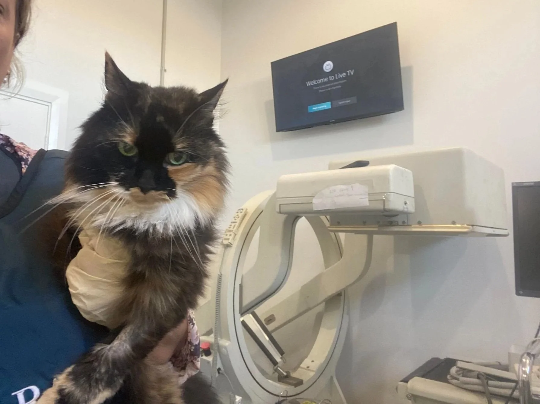
In this case, Sophia had been on medication morning and night for about 6 months with good effect. Unfortunately, many of our feline friends can be very tricky to medicate. In some cases specialised diets may be effective, however these are often not reliable, and surgery can be used for specific cases, but has the risk of significant side effects.
More recently, radioactive iodine, a treatment which can permanently cure hyperthyroidism without the need for ongoing medication, has become available for treatment in Western Australia at a small number of veterinary hospitals.
Sophia was admitted to Epsom Avenue Small Animal Clinic where she had a scan to determine the size of her thyroid. After that, a specially calculated dose of radioactive iodine was injected. Iodine in the body is quickly concentrated in the thyroid gland where its radioactivity will destroy many of the functional cells. The dose is calculated such that it will destroy just enough cells to return the thyroid to normal functioning levels.
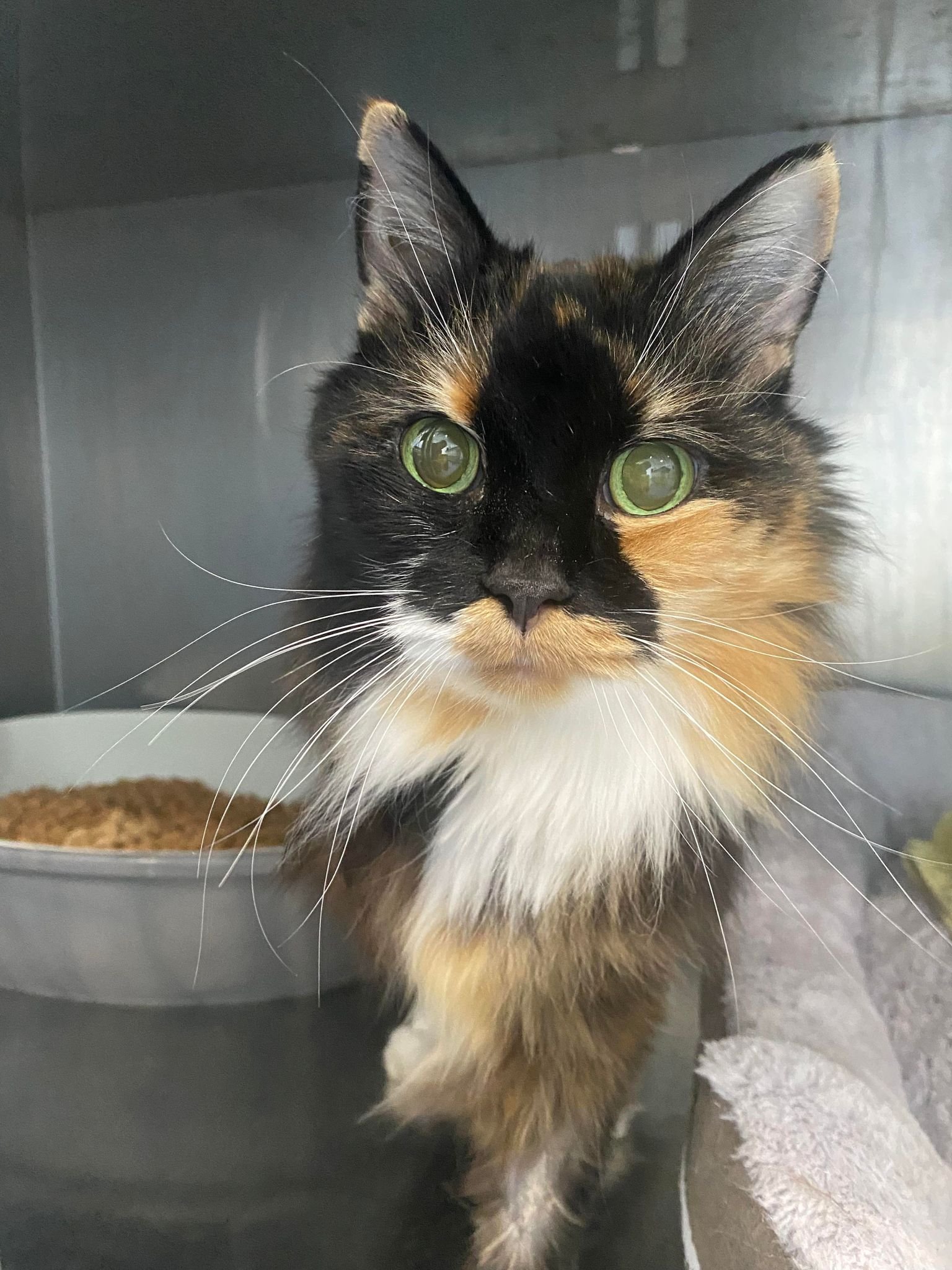
After her procedure, Sophia was required to stay in hospital for 1 week whilst radioactivity levels returned to normal, after which she was able to return home, with some extra precautionary measures for the next few weeks.
This procedure is successful in about 93% of cats. In rare instances, a small number of patients will need a second dosing. A blood test was performed 6 weeks post procedure which showed that her thyroid levels had dropped down to normal range. Another follow up blood test was then performed 6 months later, which confirmed that her thyroid is functioning at a normal level. Sophia is now free to go about her days living as a normal cat without having to deal with the stressful process of daily medications.
Franki and Her Injured Ear
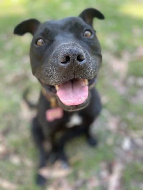
Franki is an 8 year old staffy who was bitten on the ear by another dog at the park.
Franki was initially taken to her local emergency vet where a deep laceration on her left ear was cleaned and she was started on antibiotics and anti-inflammatories.
At the time, it was decided that surgery would be difficult given the location of the wound, and that there was a good chance that it wouldn't be successful, so the wound was bandaged in the hope that it would heal without surgery.
There is often hidden damage present with dog fight wounds. The teeth crush the skin and underlying tissues, which can cause trauma and damage to the blood supply, leading to skin necrosis (tissue death) which may not be visible until several days after the initial incident.
Over the coming days, Franki's ear became more swollen and infected with some parts of the ear pinna (ear flap) becoming necrotic despite being on medication. Given the state of the ear, Dr Cindy counselled Franki’s owner that amputation of the pinna may need to occur.
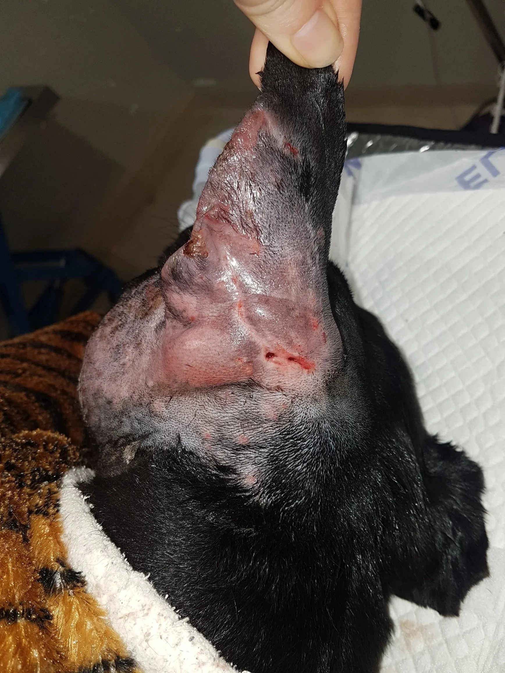
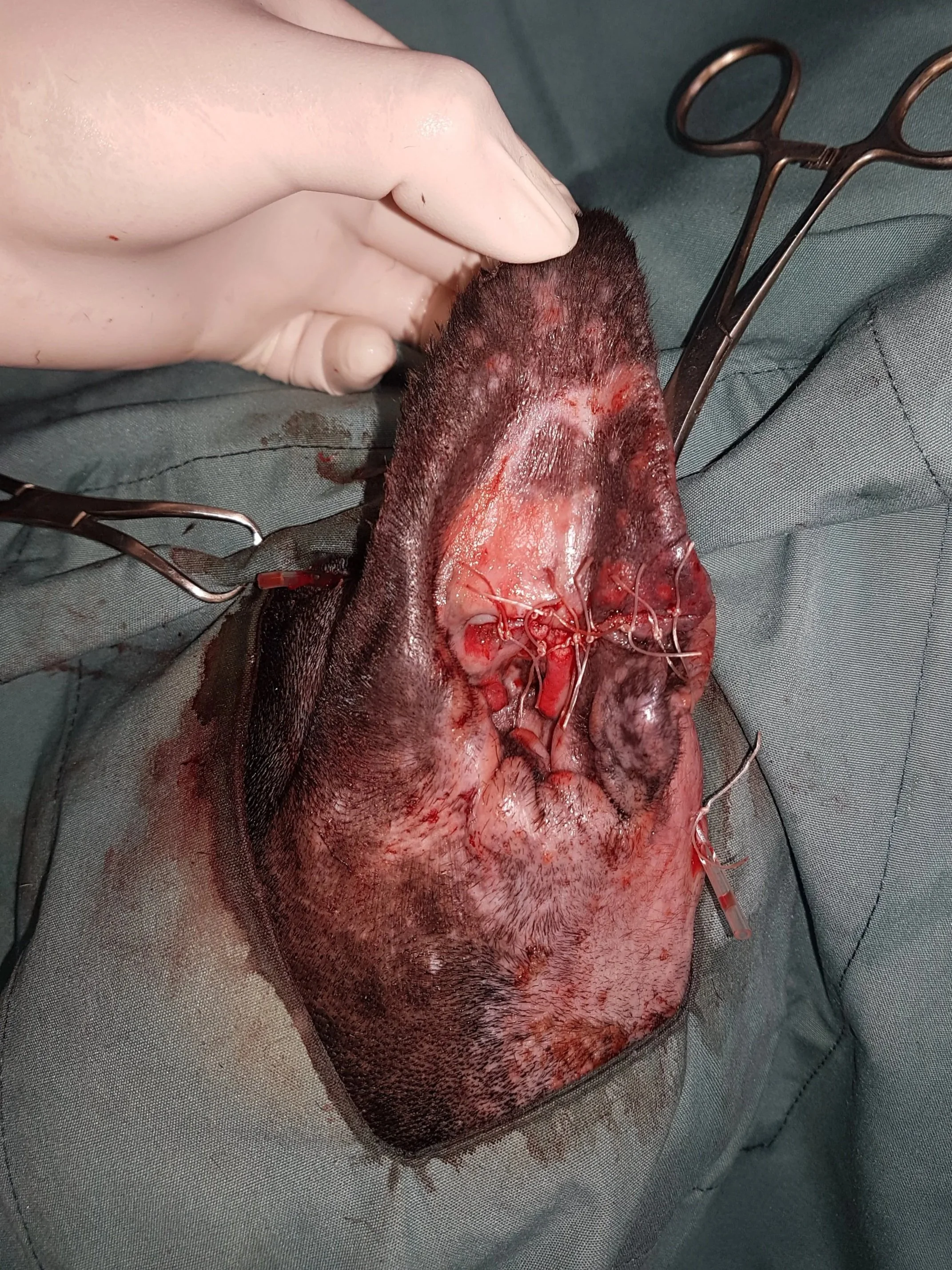
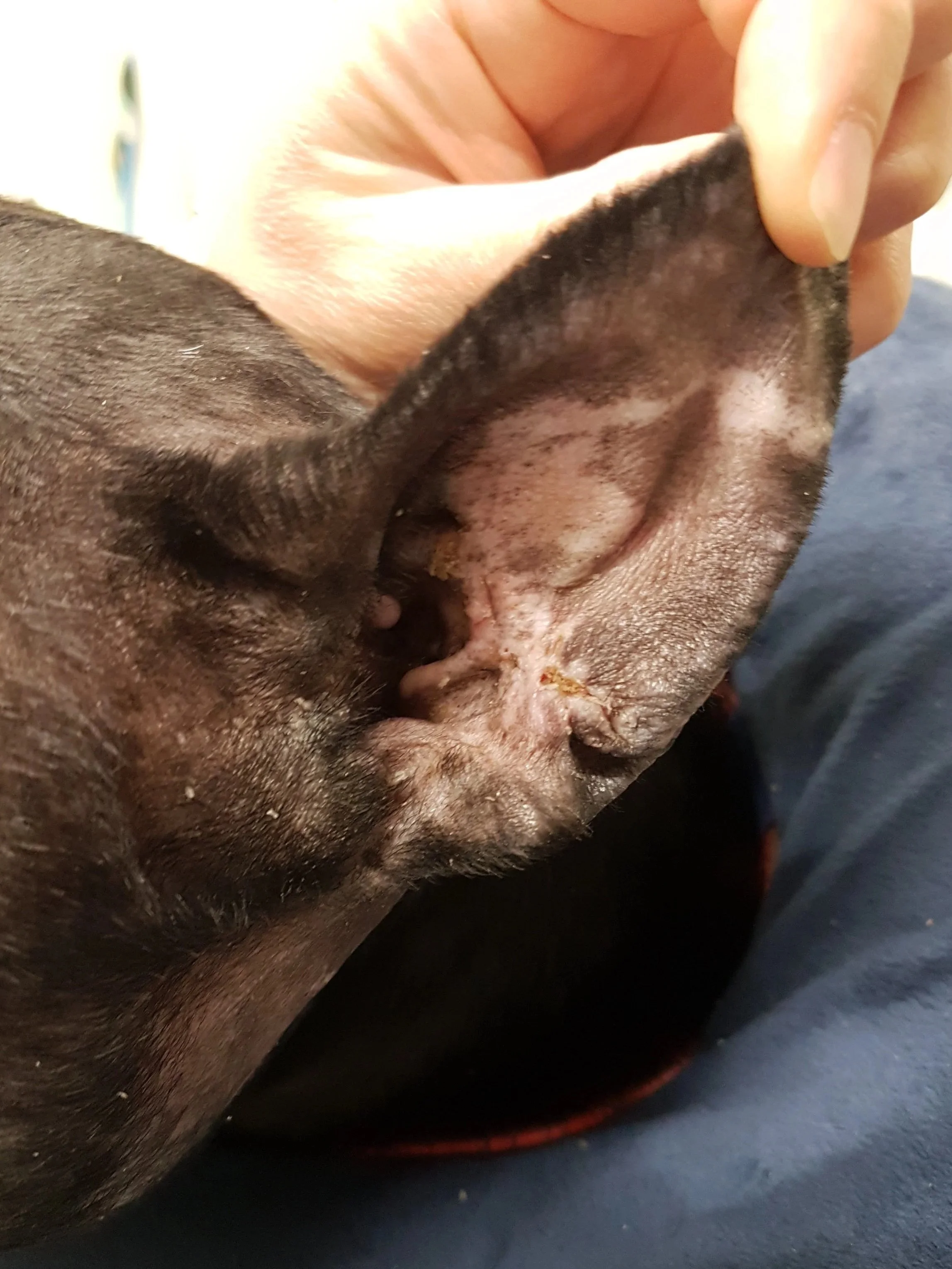
When preparing for surgery, the tip of the ear was tested, and Dr Cindy was able to confirm an intact blood supply which meant that the ear could likely be saved. One potential drawback to surgical repair of the ear however, is that the edges of the repaired tissue may contract, deforming the ear pinna, but this is still generally preferable to amputation.
During surgery, large amounts of infected material and pus were cleaned away, necrotic tissue surgically removed, and a sample was sent to an external laboratory to culture the bacteria. The ear pinna was sutured back together.
2 weeks later, she had healed with minimal deformity to the ear.
Ollie's Park Trip
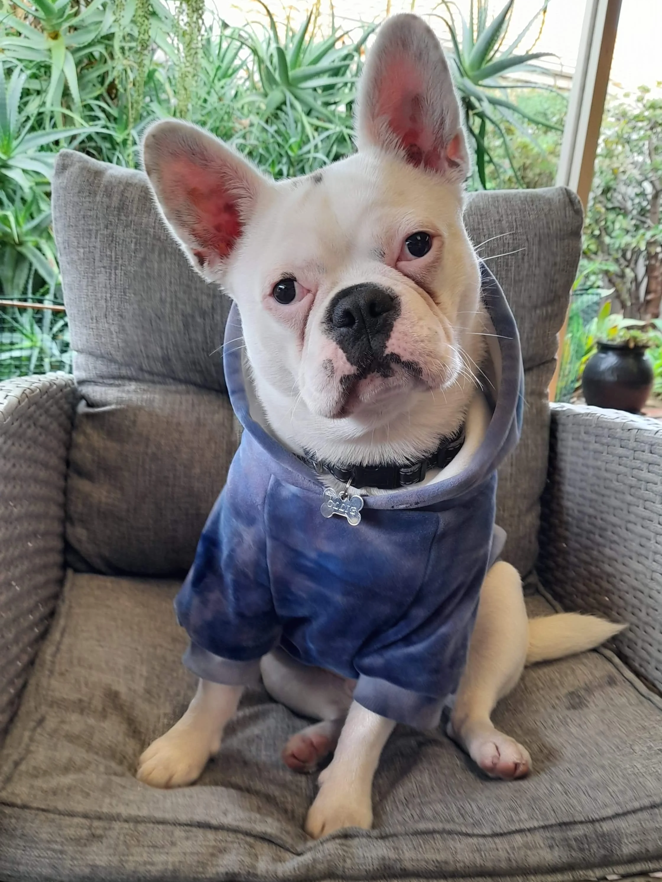
Ollie is a 1yr old French Bulldog X Pug who presented to us as he suddenly became very unwell, wobbly on his feet and hyper-reactive to noises following a visit to the park. On examination, Ollie seemed very disoriented and was constantly swaying and unable to maintain balance. His pupils were miotic (constricted) and did not respond appropriately to light. His body temperature was also very low.
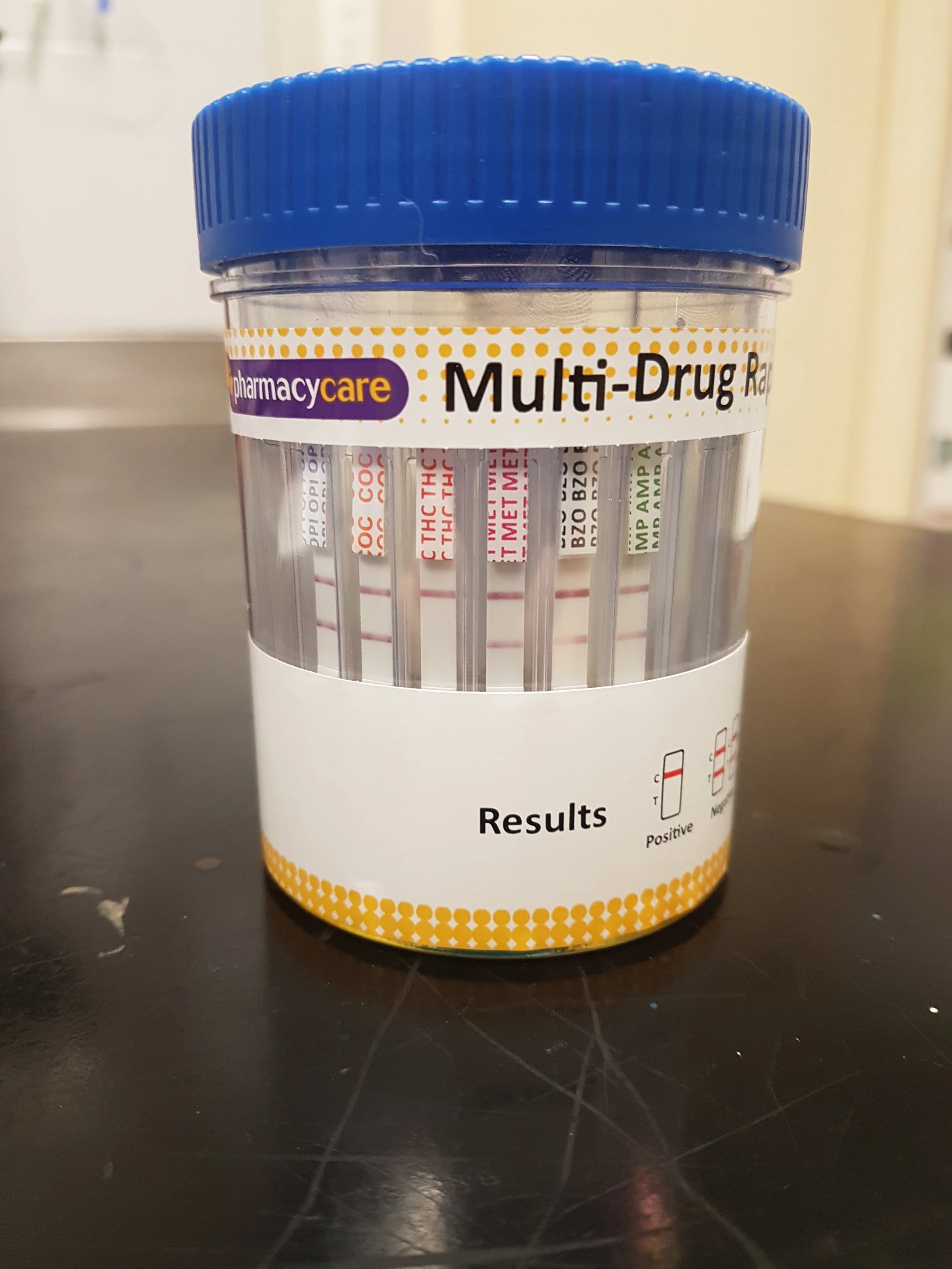
Ollie was admitted into hospital for further investigation and supportive care. He was started on intravenous fluid therapy in case of possible toxin exposure, as fluids can help flush the toxins out of the system quicker. Additionally, bloodwork was performed, which showed mild inflammatory changes. There were concerns that Ollie may have accidentally ingested a toxin or recreational drug, and so drug tests were performed on his urine as well. Marijuana was detected in Ollie’s urine, and this was likely responsible for the clinical signs seen.
Marijuana intoxication can occur in any dog that accidentally ingests the substance, and symptoms typically begin 30-90mins following consumption. The typical signs include incoordination, slow heart rate, altered pupil size (usually dilated), sedation and occasionally urinary incontinence. In severe cases, vomiting, seizures, and heightened sensitivity to noise and/or light can occur.
For Ollie, he was administered activated charcoal, which is commonly used in the treatment of poisonings. Activated charcoal is given orally and binds to toxins as it passes through the body. Additionally, Ollie was given a dose of Naloxone intravenously. Naloxone is a medication used to reverse the effects of opioid drugs, but has been reported to be successful in the treatment of cannabis overdose.
Ollie was monitored closely in hospital, and began to show significant improvement as the day went on. Just several hours following his initial presentation, Ollie was able to walk normally again with his tail wagging, and was no longer hyper-reactive to noises. He was able to be reunited with his family that evening.
Ollie was very fortunate that his owners picked up that something was wrong early, and brought him in to see us so that supportive treatment could be started. Whilst the chances of fatality through marajuana overdose is small, it is still possible in our companion animals. We contacted Ollie’s owners the next day to check in on him, and we were happy to hear that Ollie was back to his usual crazy self!
Bruiser And His Treacherous Treat
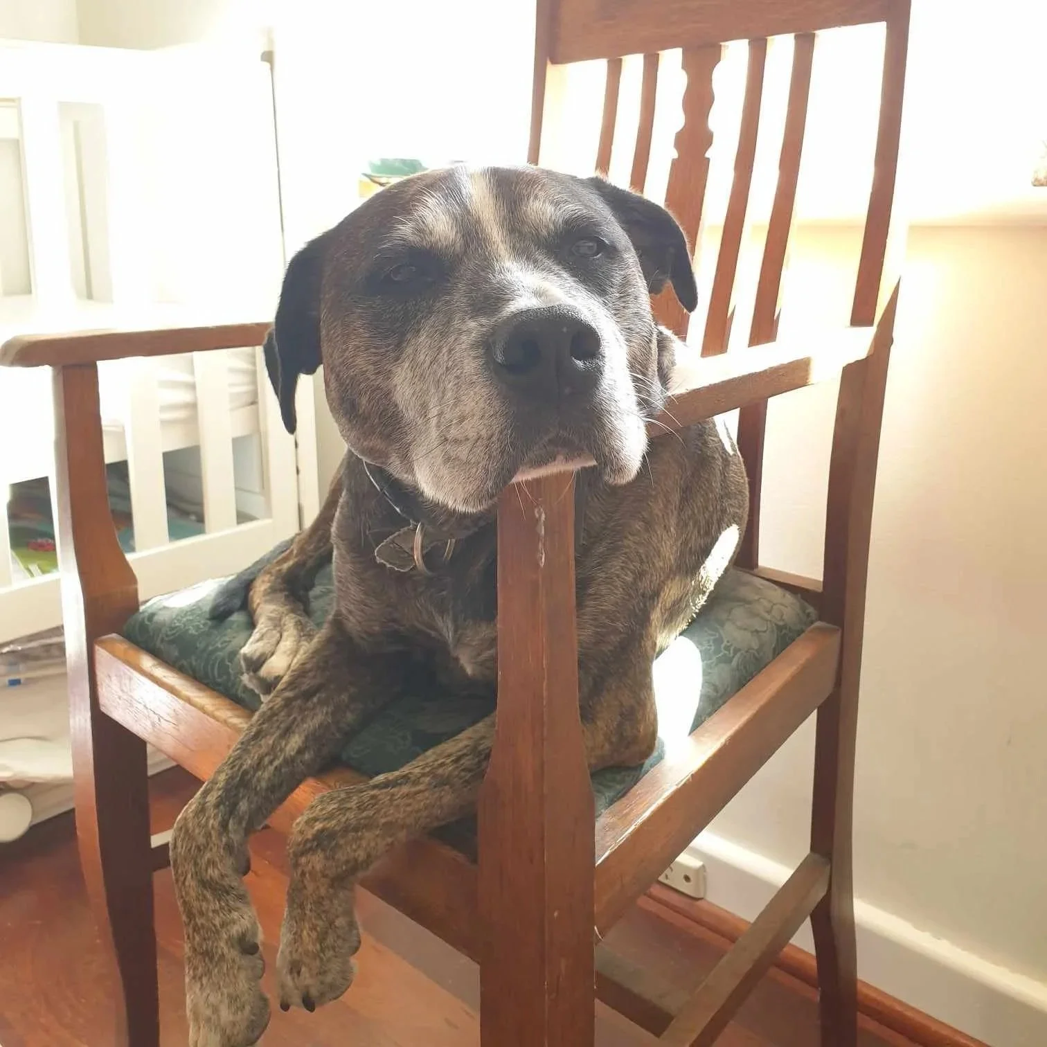
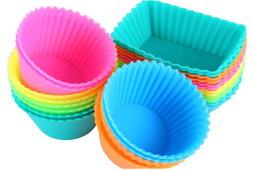
Bruiser is a 7 year old Staffy cross who came to us after an ill fated snack. Bruiser managed to get a hold of a silicone cupcake mould containing half of a cupcake which he promptly swallowed whole before his people could stop him.
Plastic or rubber food packaging which still contains remnants of food are often targeted by our canine friends as they have the same aroma and taste as the food they contained, and dogs will sometimes swallow the packaging whole before they are able to be stopped. Meat skewers (with or without meat attached) are also occasional targets for the same reason.
Whilst the dangers of swallowing a skewer whole are obvious, the dangers associated with swallowing soft plastic or silicone may be less apparent. The digestive tract is unable to break these products down, and once in the stomach, the only way to go is forward, through the narrow intestines. This will often lead to blockages in the gastrointestinal tract, preventing food and other digestive material from passing through causing severe vomiting.
More critically, pressure caused by the blockage can prevent blood flow to a portion of the intestine, which will result in tissue death and ultimately leading to break-down of the intestines, causing the abdomen to become septic. Without immediate treatment, this will result in death.
Because of this danger, it is imperative to remove the material before it can move into the intestine and cause a blockage. There are several ways to achieve this.
Method 1 is to make the patient vomit up the material. This is the least invasive method and can often be an easy way to solve the issue, as long as there's no risk of the material causing damage on the way up. It does have its limitations however. In this case, Bruiser was given apomorphine, a medication which rapidly causes a short duration of significant vomiting. Unfortunately, all he was able to bring up was some foamy liquid as the silicone cupcake mould was blocking the outlet of his stomach.
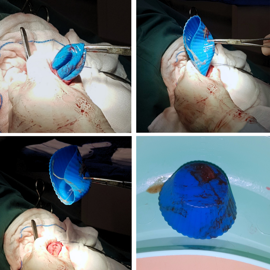
Method 2 involves a general anaesthetic to feed a scope, fitted with a small grabbing claw, down the oesophagus. Unfortunately in this case, the slippery nature of the silicone, coupled with the rounded edges made it impossible to properly grip the mould for extraction.
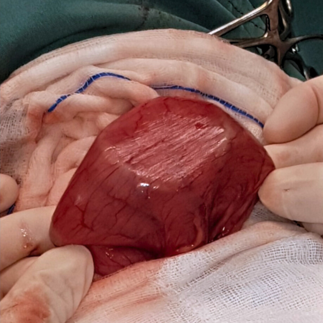
Method 3 is to surgically open up the stomach to manually remove the foreign object. This is the most invasive method, however in many cases it is the only viable option. Bruiser went into surgery that same day. A large incision was made into his abdomen and then an incision was made into his stomach, where the silicone mould was removed.
Following his surgery, Bruiser was a bit groggy for a few days, but soon recovered and was back to his usual self.
Bruiser's people would like to share his story to hopefully prevent this from happening to another unsuspecting pooch.
Apollo’s Dangerous Snack
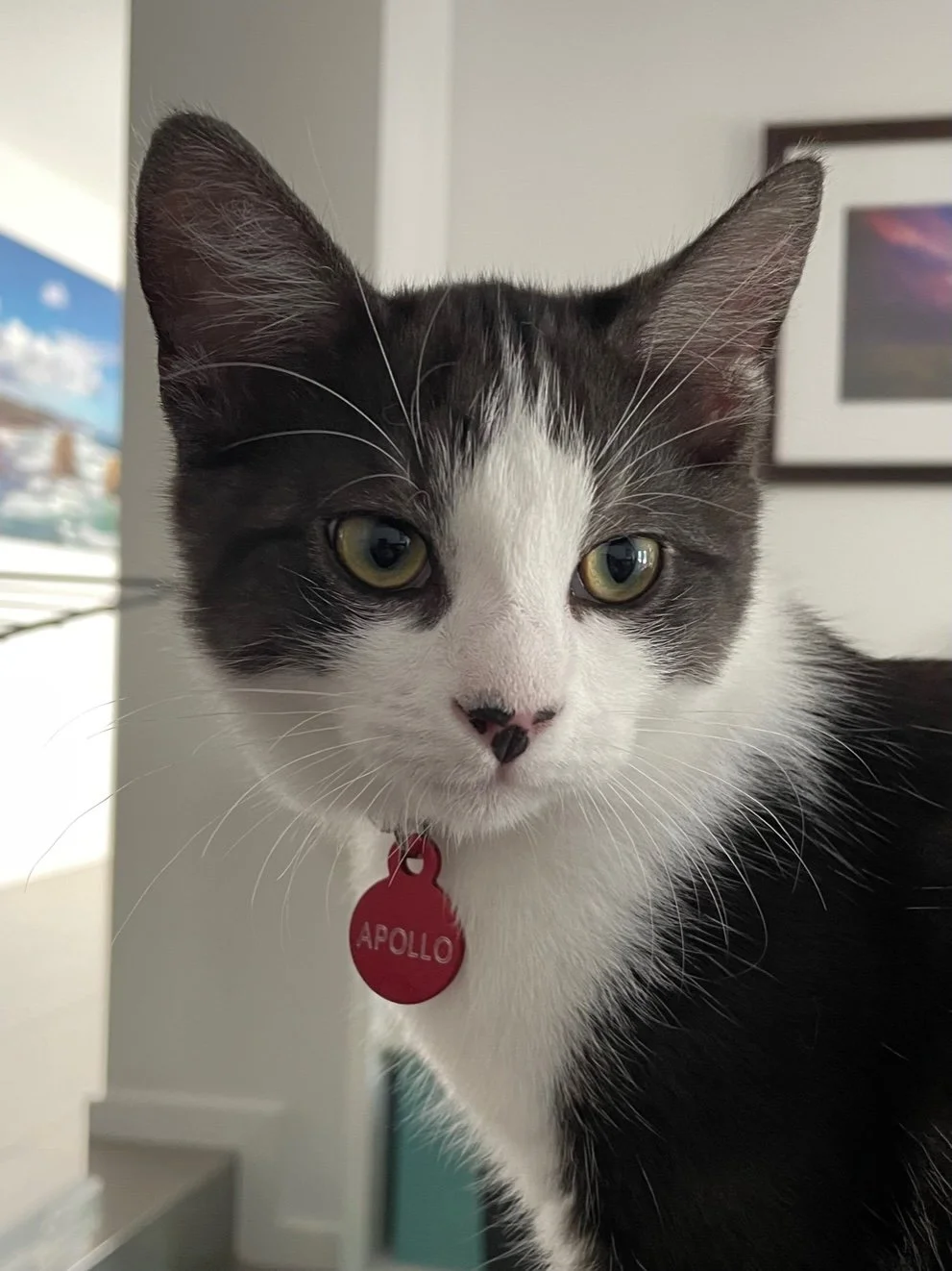
Apollo, a 9.5m old kitten, presented to KSAH because he had been off food for a few days. His owners had seen him chew and possibly swallow a piece of ribbon or elastic from a toy before he became unwell.
Apollo was repeatedly uncomfortable (hissing and trying to bite) when we palpated the front half of his abdomen. Due to his abdominal pain and history of potentially ingesting something he shouldn’t have, his owners decided to proceed with abdominal ultrasound the following day.
The ultrasound scan showed that Apollo had a foreign object in his stomach. A type of endoscopy called gastroscopy was performed under general anaesthetic to try to retrieve the foreign object from his stomach. A gastroscope involves passing a camera down the oesophagus into the stomach to visually assess what is present and if appropriate, pass a pair of forceps down the endoscope tubing to try to grab the object. This means that surgery can be avoided.
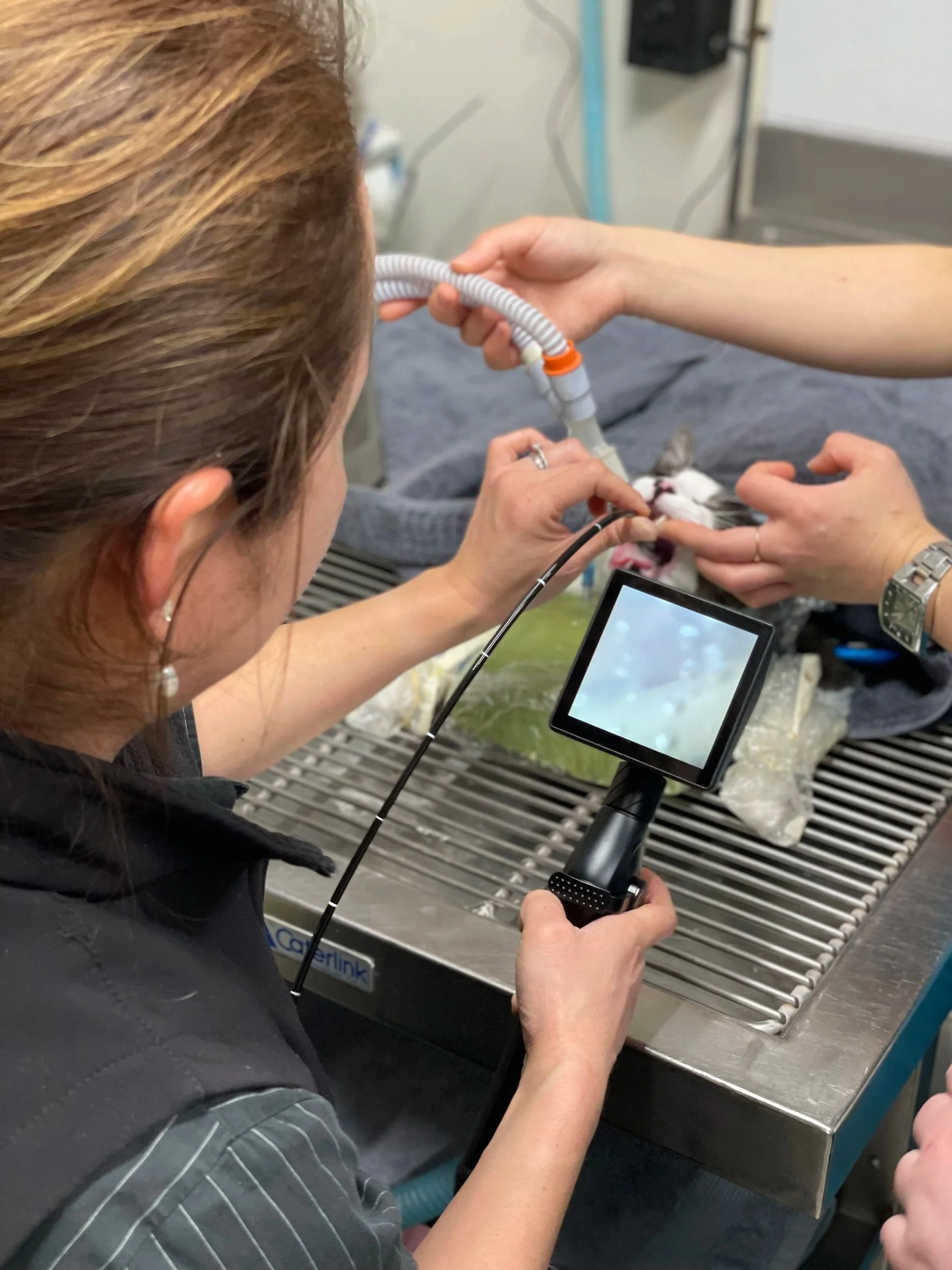
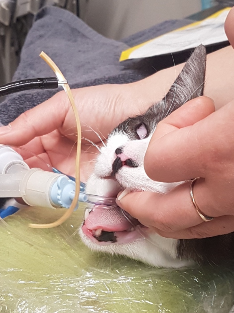
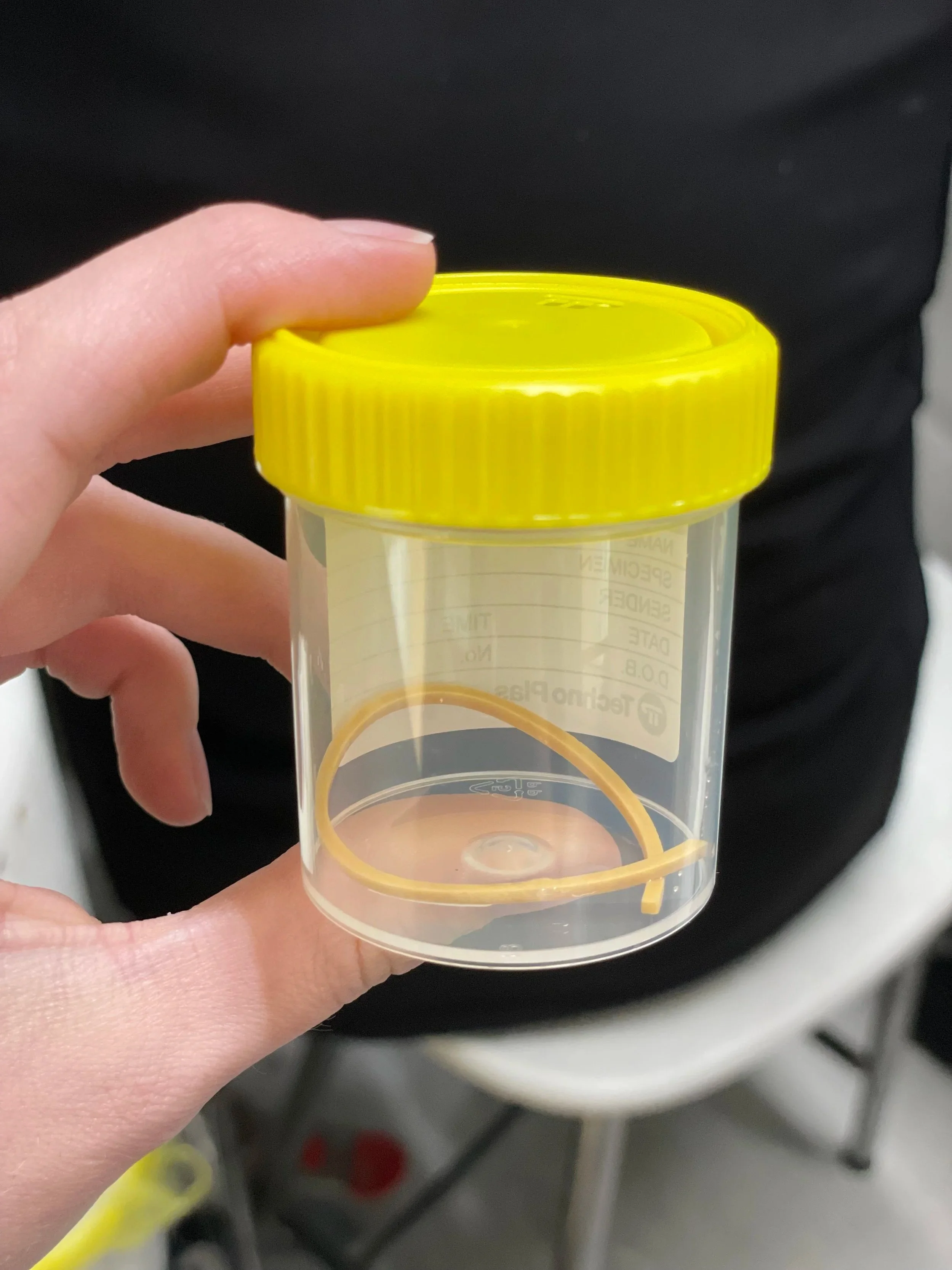
Dr Cindy commenced the gastroscopy with assistance from our nursing team and upon entering the stomach, identified a rubber band floating freely in his stomach! The foreign body forceps were passed through the endoscope and a broken elastic band was successfully removed from the stomach. The endoscope was advanced back into the stomach to ensure there were no other objects present.
After waking up from anaesthetic Apollo’s stomach was much more comfortable and he had a very good appetite that evening once he got home. He continued to recover uneventfully over the following days and was back to his mischievous self in no time.
Any foreign object can cause an obstruction (blockage) of the stomach or intestines depending on the size of the object/patient (that then requires surgical removal), but string-like items such as elastic bands, string, fabric and rope can cause severe intestinal damage (e.g. perforation) that can result in lengths of intestine being removed.
It is important that if you ever notice your animal eating something they are not supposed to, that you contact your veterinary team for advice and treatment as soon as possible.
Ben's Year Long Macadamia Snack
Ben is a 7 year old kelpie cross who was presented to KSAH for vomiting. He had been at an emergency clinic the night before and was not improving, so his owners brought him in for further investigation.
He was flat and lethargic and had vomited up bloody fluid. 12 months ago Ben had managed to raid a bowl of macadamia nuts (some of which were still in their shells) and since then has been occasionally vomiting up pieces of indigestible macadamia shells. We were concerned that one of these nuts had become lodged in his intestines and caused a serious obstruction.
Ben was admitted for abdominal ultrasound which identified that there was a 2.5cm round foreign object in his jejunum (middle part of his small intestine) and that the surrounding tissues were very inflamed. Dr Cindy then proceeded to surgery with Ben as it was very unlikely that the macadamia nut would pass on its own and there was a risk that his intestines would rupture if the object was left in place.
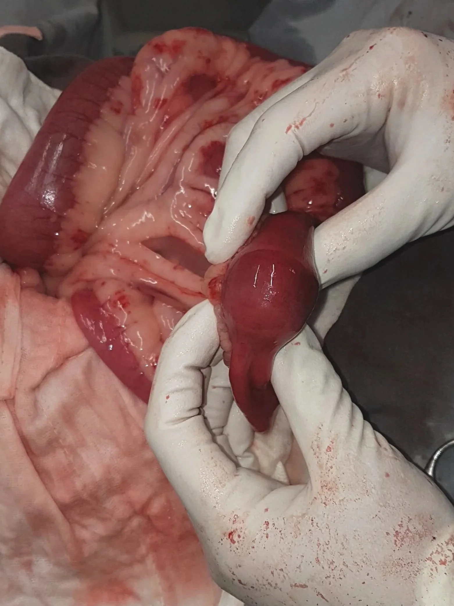
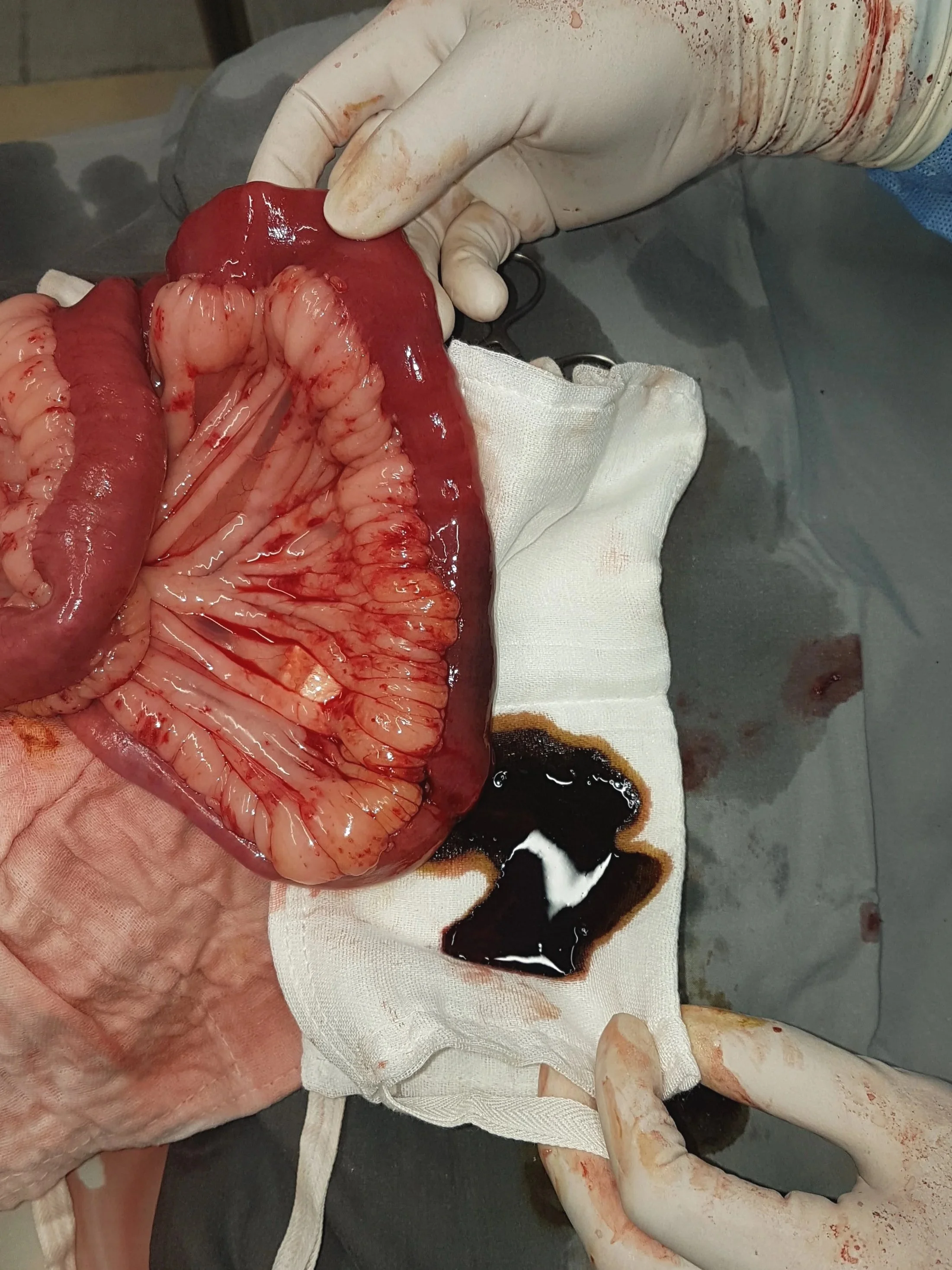
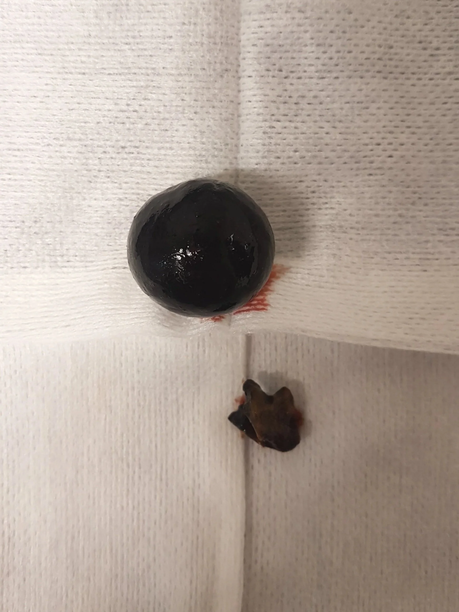
During surgery, an 8cm section of intestine behind the foreign body was very dark (should normally be a healthy pink) and it had likely died due to poor blood flow. An incision was made over the foreign body exposing an intact macadamia nut and an additional partial shell.
Due to the severely diseased section of intestine and poor intestinal contractions on either side of this area (indicating that the intestines had been badly damaged despite looking healthy), a total of 28.5cm of Ben’s intestine had to be removed. A procedure called an end-to-end anastomosis was performed to reattach the two ends of his intestine back together after the unhealthy portion had been removed.
Ben was discharged the following day after spending the night at emergency for supportive care and was monitored closely at home by his family for any complications.
Ben steadily improved over the days to come and is now back to his bright and happy self (and hopefully not snacking on inappropriate things).
Brodey's Tongue Tied
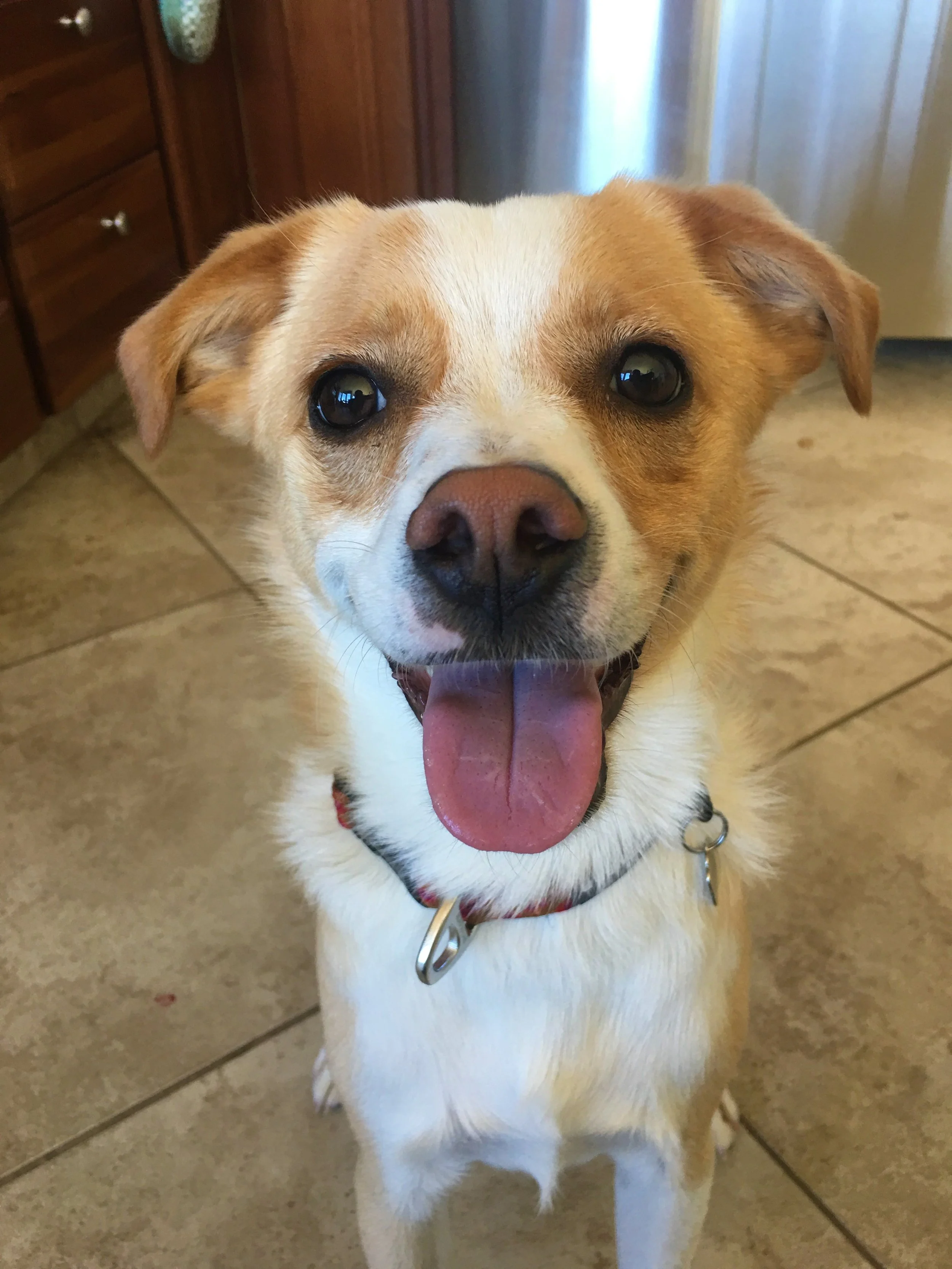
Brodey is a 2 year old Jack Russell Terrier X who presented to us for gagging whenever he tried to eat.
Despite some anti-inflammatory medication to treat a suspected throat irritation, Brodey continued gagging intermittently.
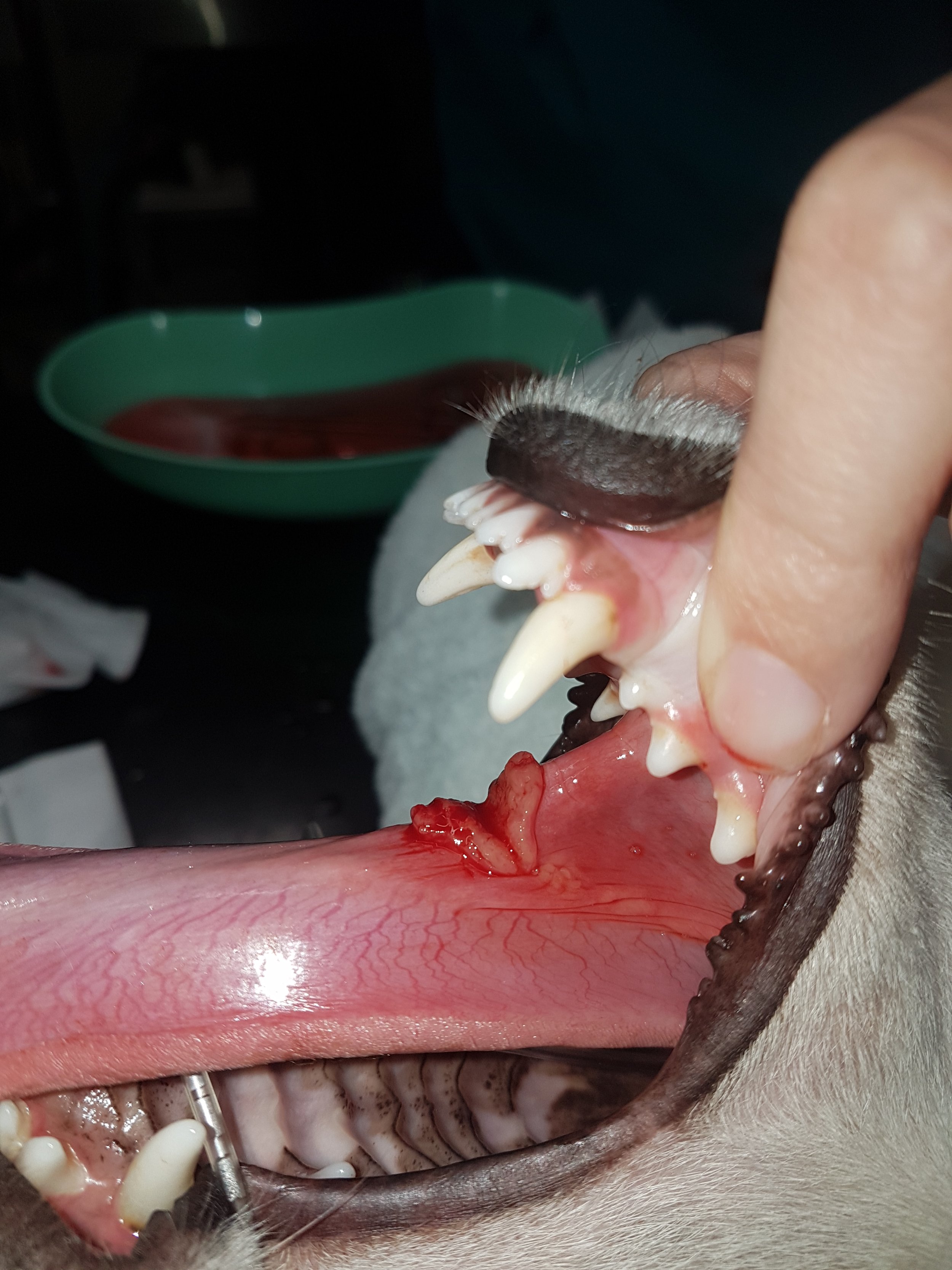
Dr Sam anaesthetised Brodey to examine his throat in further detail. He noticed a small white thread was looped around the base of Brodey’s tongue, and it continued down into the oesophagus.
The thread had caused significant trauma to the underside of the tongue.
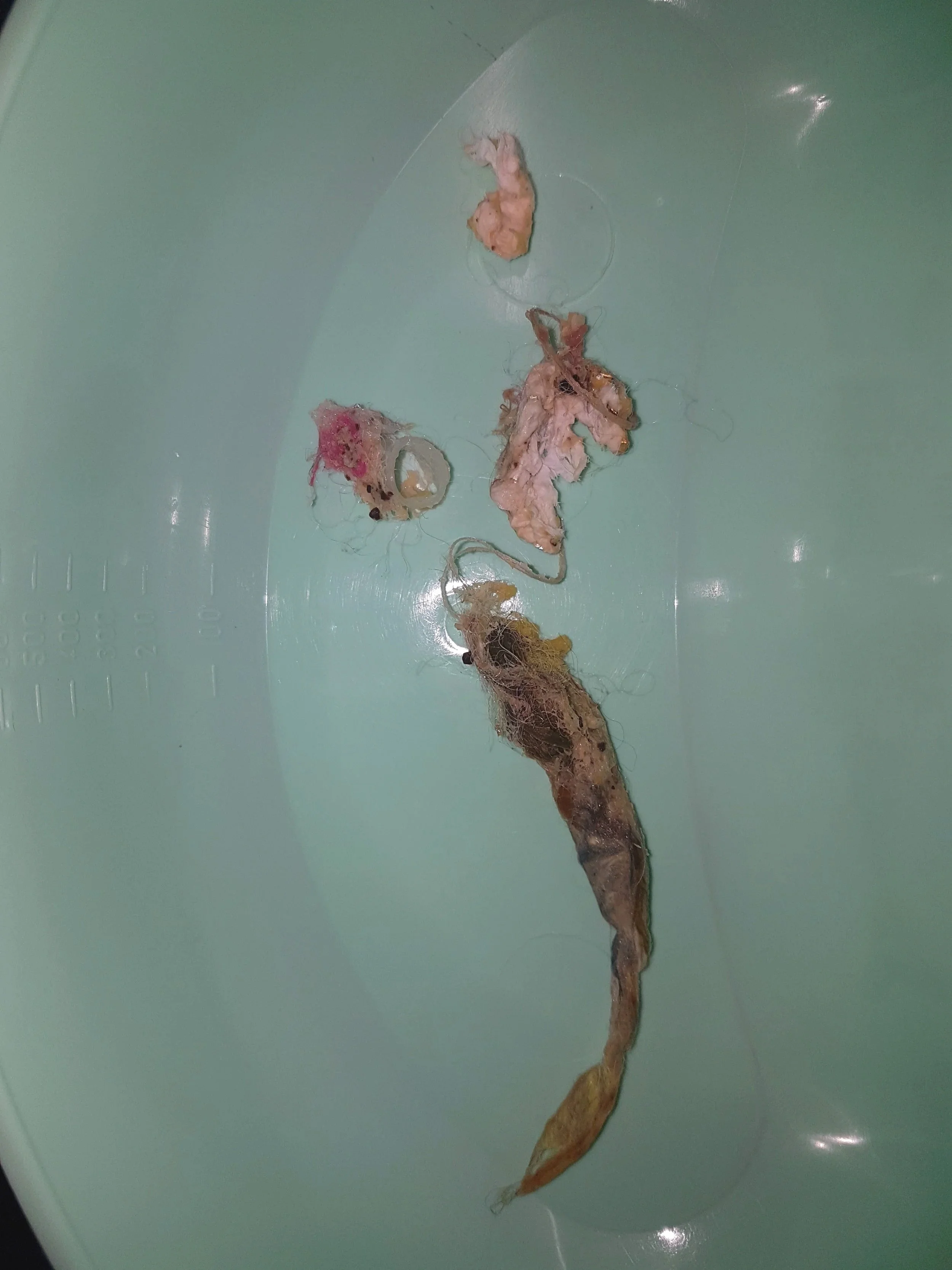
When it was pulled from the oesophagus, it was discovered that a large amount of hair, fur and foreign material was attached to the end of the thread.
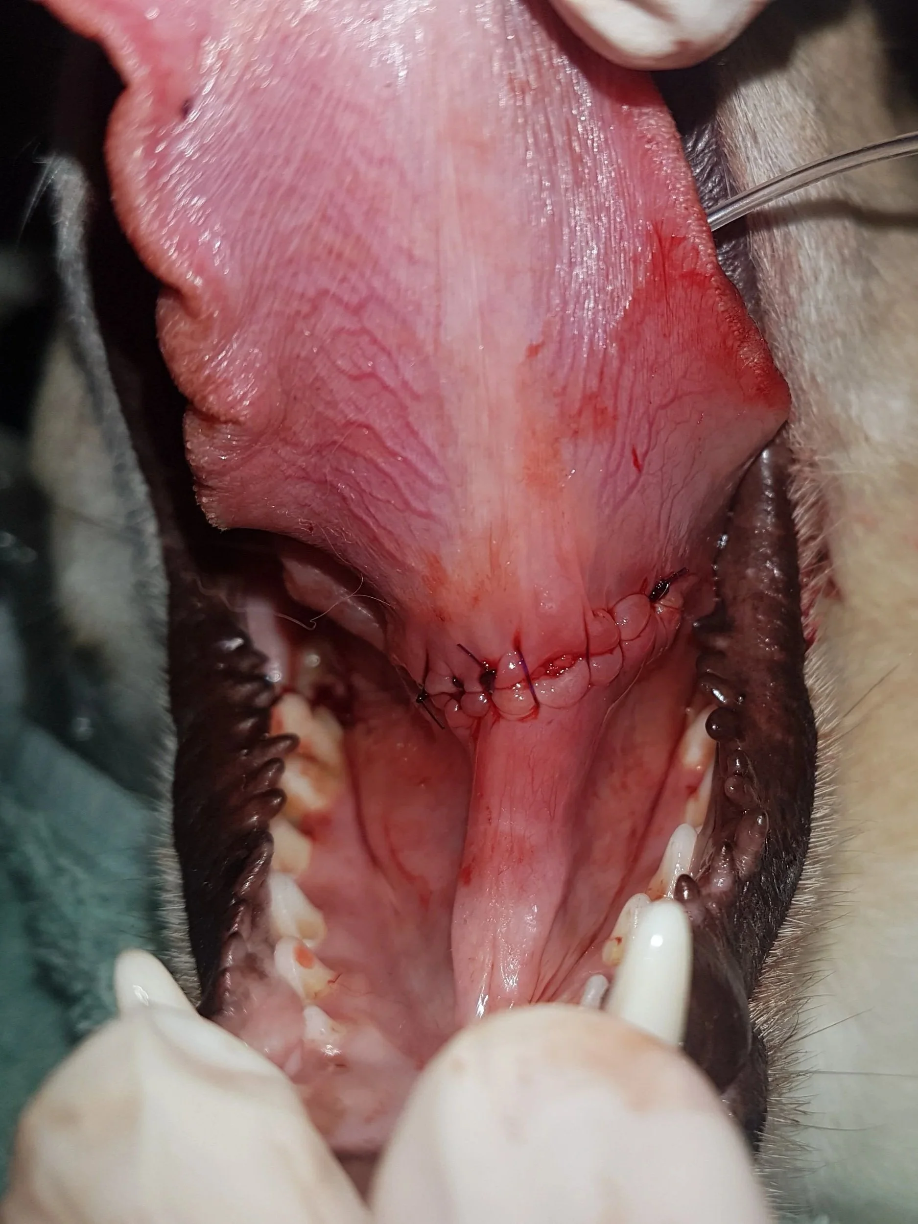
The laceration to the tongue was surgically debrided (surgical removal of necrotic tissue) and repaired, and Brodey was sent home with a course of pain relief and antibiotics.
He has since made a good recovery, and the gagging has completely resolved after this procedure.
Bridget and Her Sore Eye
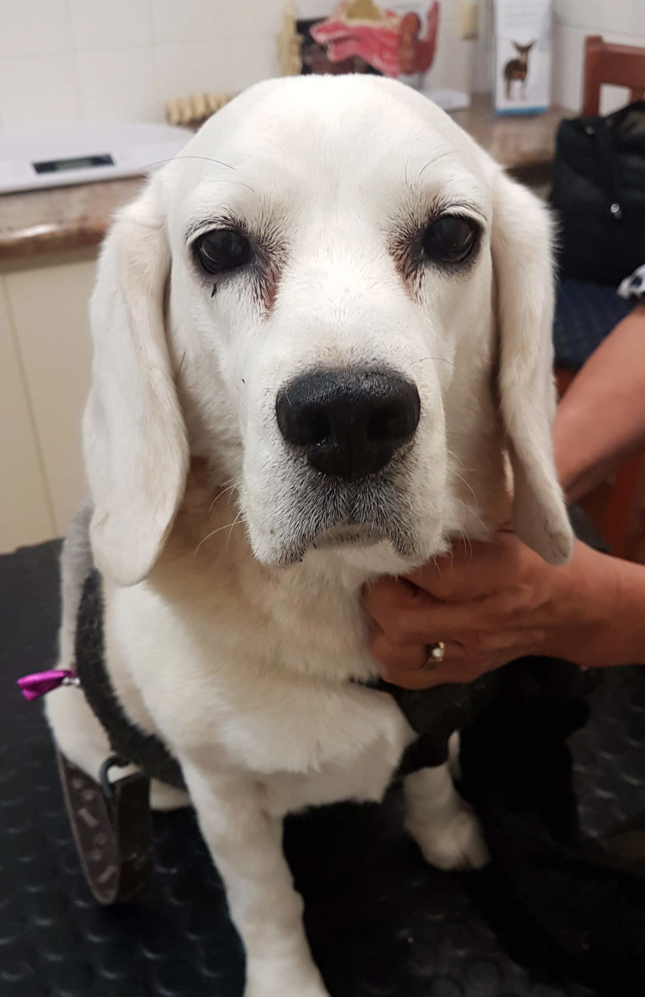
Bridget, a 12 year old Beagle, came to us with a weepy right eye.
Dr Cindy could see a 2mm foreign object in the right eye. The sclera of the eye (the dense, white, fibrous membrane that forms part of the external covering of the eye) was very inflamed and irritated. It was recommended that the foreign material be removed under general anaesthetic, and Bridget’s dedicated owners went ahead with the procedure the very next day.
Prior to anaesthetising Bridget, her intraocular pressures (the pressure within the eyeballs) were measured to ensure that they were within normal limits. Dr Jess then proceeded to remove the foreign object from Bridget’s right eye.
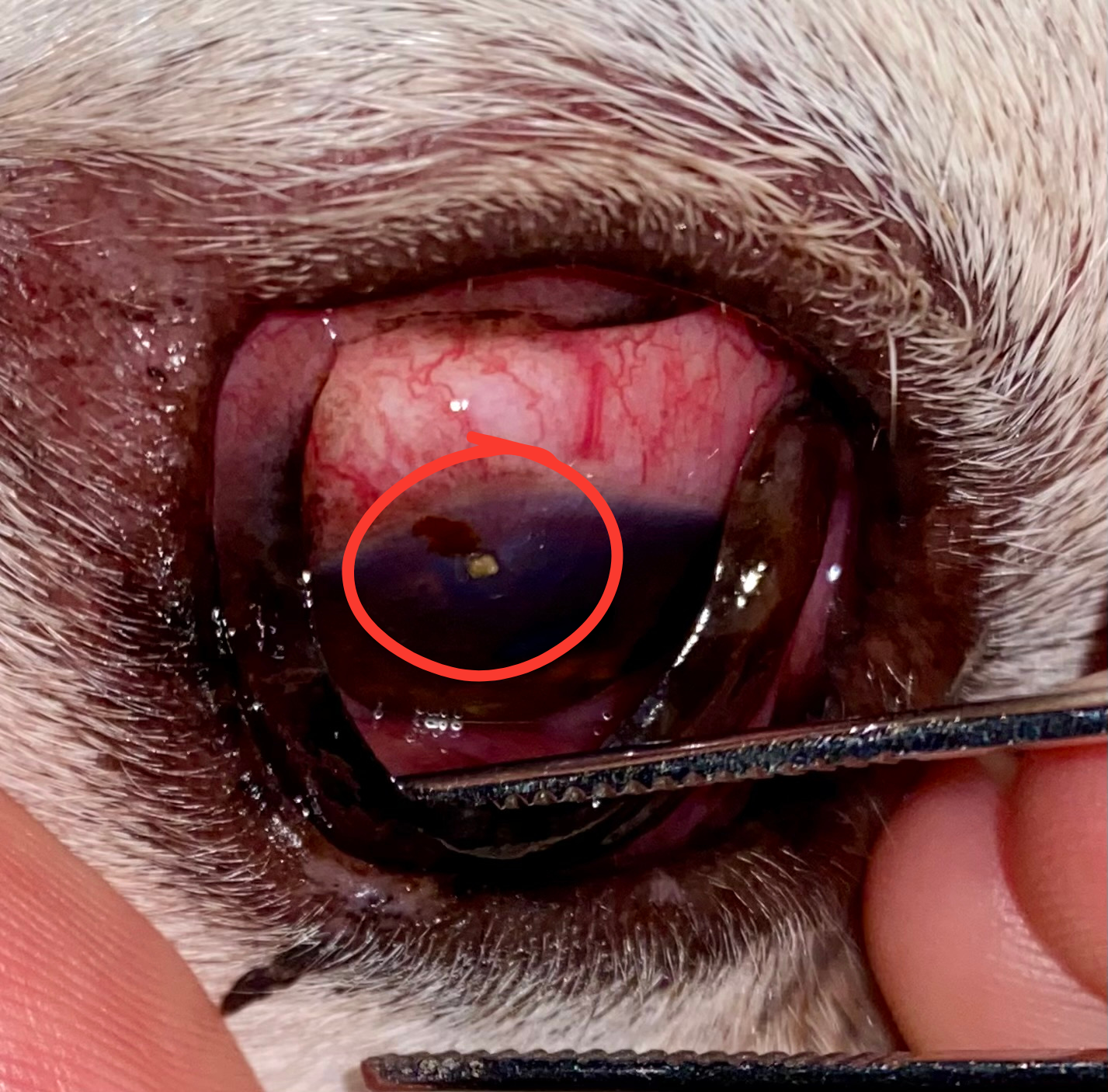
This was done via applying very gentle pressure and hooking out the foreign material with the tip of a very fine needle. A lot of care had to be taken to not inadvertently cause more damage to the eye. An approximately 1.5mm wide x 1mm deep defect to the cornea was left behind from the offending object, which healed without issues via a course of antibacterial eye drops and pain relief post operatively.
It is important to remember that because eyes are such fragile yet important structures to the body, if you ever notice that your pet’s eyes just ‘don’t seem quite right’, do give us a call for a chat and further advice.
Bella's Pyometra
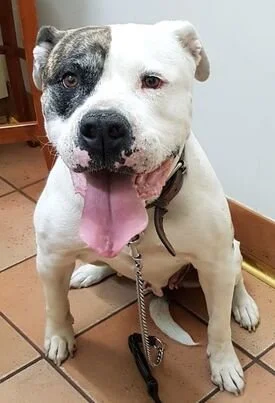
Bella is a seven and a half year old American Bulldog that presented to KSAH as she was losing weight, and drinking and urinating more. At the time of presentation Bella was an entire female and had her last heat 6 weeks prior. On physical exam she was underweight and had multiple masses adjacent and attached to her mammary tissue.
A full blood panel was conducted and showed evidence of chronic (long term) inflammation and anaemia (decrease in red blood cells). Dr Cindy suspected Bella had either cancer in her mammary chain, pyometra, or potentially both.
Pyometra is a life-threatening infection of the uterus that can affect female dogs and cats that have not been desexed. It occurs as a result of hormonal changes in the reproductive tract that cause thickening of the uterine wall in preparation for pregnancy. If no pregnancy occurs after consecutive heat cycles, the lining continues to thicken and creates an ideal environment for bacterial infection to occur.
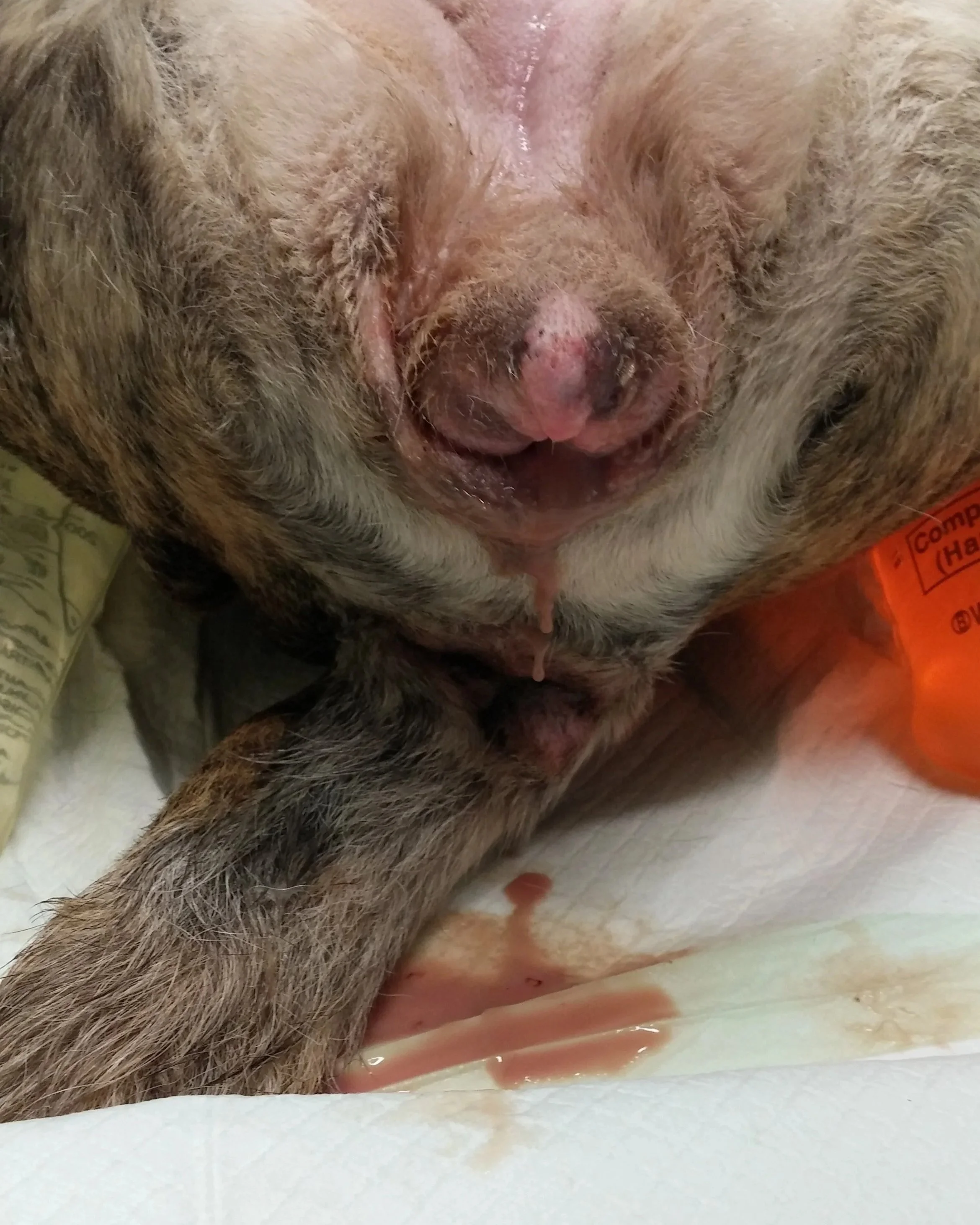
Surgery is always indicated to remove the ovaries and infected uterus. Pyometra can be classed as “open” (pus discharge draining from the vulva) or “closed” (pus is enclosed within the uterus without visible discharge from the vulva).
Bella initially had a closed pyometra, which eventually progressed to a partially open pyometra and was draining green pus.
An ultrasound was conducted and a diagnosis of pyometra was confirmed. Bella was placed on aggressive fluid therapy, administered intravenous antibiotics and prepared for surgery. As her health condition was poor, general anaesthetic was very risky for her. She was closely monitored and supported by our skilled team of nurses throughout her procedure.
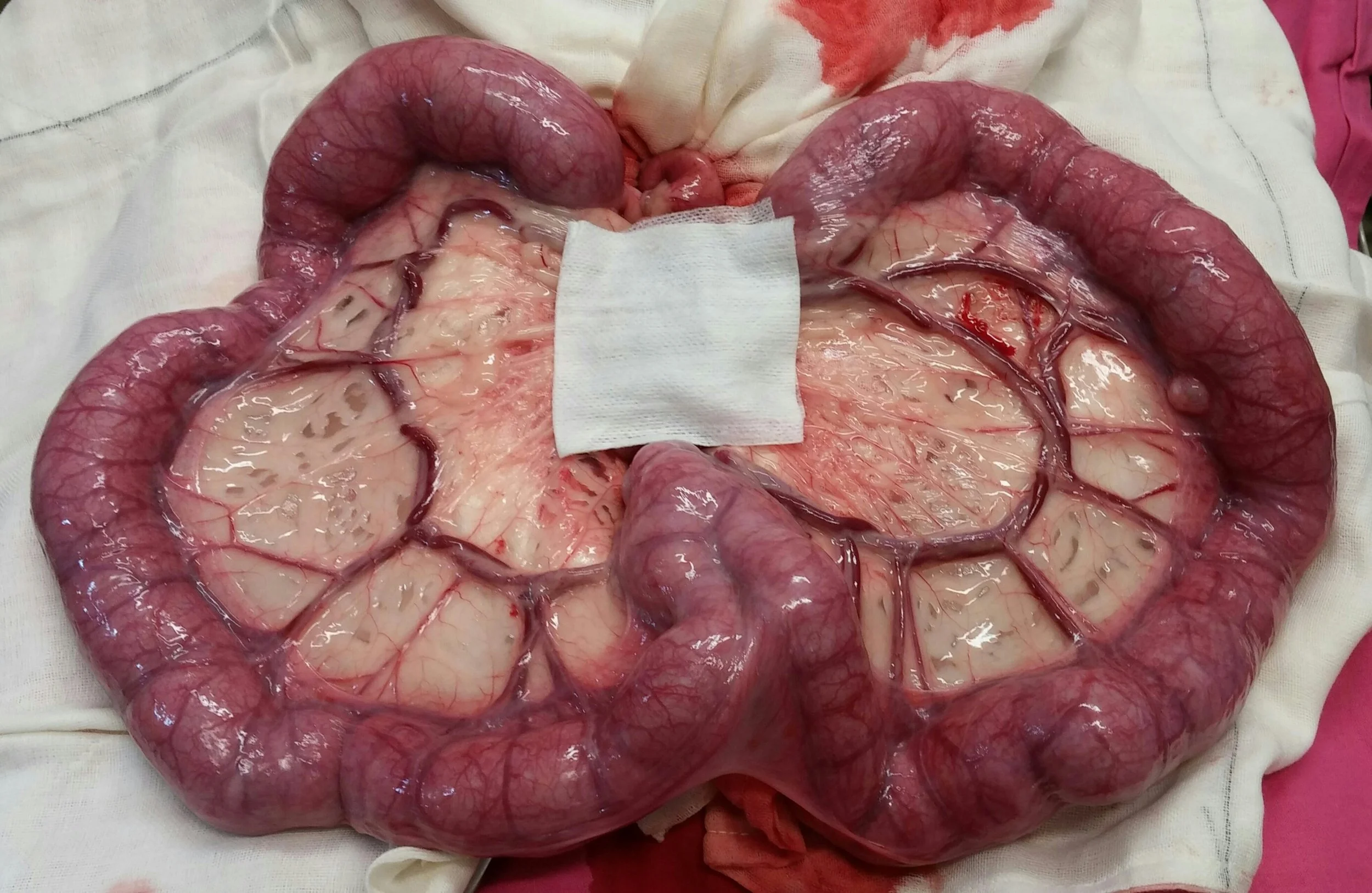
During surgery an extremely enlarged, inflamed and pus-filled uterus was identified and removed by Dr Cindy. In the photo on the left, the uterus weighed 1.2kg and is compared to a healthy uterus of a similar sized dog. A 7.5cm x 7.5cm gauze swab was placed next to each uterus for size comparison.
Her owner is very happy that Bella has since recovered well from her surgical procedure.
Tom's Sewing Together His 9 Lives
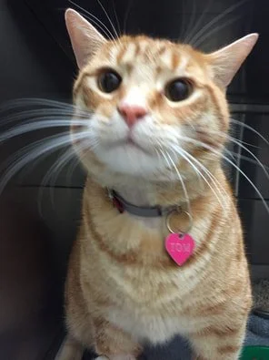
Meet Tom, he's a mischievous three year old cat who got into a spot of bother when he managed to swallow something very worrisome! Tom's owner brought him into Karrinyup Small Animal Hospital after they noticed that he had eaten a long piece of thread. Since then he had vomited and was gagging repeatedly. He was also very lethargic and was off his food.
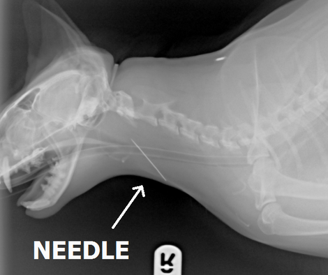
Tom was admitted into hospital and x-rays showed he had a large, long metal object lodged in his throat. The foreign body identified was a sewing needle and can be clearly seen in the x-ray below.
While Tom was under general anaesthetic, Dr. Ruth was able to pass an endoscope down into his throat to find the needle in question. Using long forceps, the needle (which also had some thread still attached) was removed from poor Tom's throat.
Tom stayed in hospital for a few days on antibiotics, anti-inflammatories and pain relief. Within a few days he was back to normal at home and back to his old tricks again (but staying away from needles and thread!)
Tom was very lucky that the needle did not cause more extensive damage to his throat, oesophagous and trachea. Thread and other string-like objects can also be very dangerous for cats, causing what is known as a “linear foreign body”. These lodge in the intestines and cause it to bunch up and cause extensive intestinal damage requiring emergency surgery. It's best to keep objects like these well away from cats.
Alfie and His Unwanted Passengers
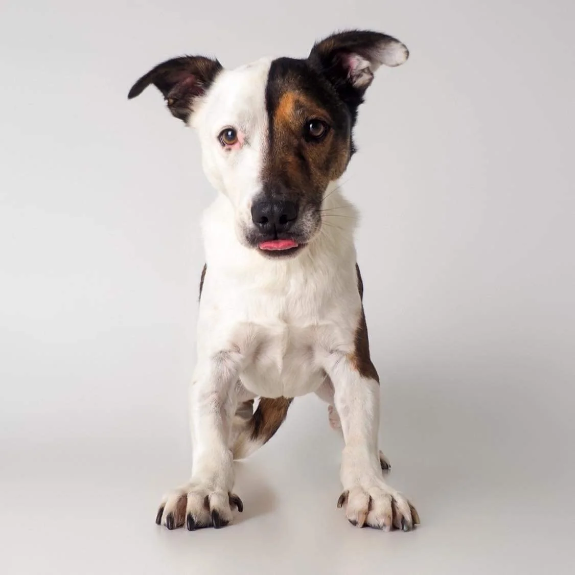
This is adorable Alfie. He's a one year old Border Collie x Staffy. Young Alfie was found
as a stray by the City of Stirling Rangers and was brought into Karrinyup Small Animal Hospital for veterinary assessment.
Poor Alfie was in a very sorry state when he was brought into us. He had a nasty skin infection and a lot fur loss around his face, under his neck and all over his body. He was very itchy and sore. A skin scrape revealed that Alfie had a skin mite known as “Demodex”.
Demodex mites are microscopic and live deep in the hair follicles of the skin. They are not transmissible to other animals or humans. In low numbers, Demodex can be a normal inhabitant of dog skin.
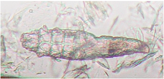
In some cases, however, these mites can take over and lead to a condition known as “Demodecosis”. This can happen quite commonly in puppies and young dogs less than 2 years of age. It is usually due to their growing/immature immune system allowing the mite to proliferate.
Despite being very sore, Alfie had the most beautiful nature in hospital. He was very brave when having his skin sores cleaned and treated. Alfie was started on antibiotics and anti-inflammatories to treat his skin infection. He was also started on an anti-parasitic medication to treat the Demodex mite.
Alfie's loving and trusting nature won over the hearts of all of the vets and nurses at Karrinyup Small Animal Hospital. So much so, that one of our nurses decided to adopt Alfie and give him the loving forever home he deserves.
We are pleased to say that Alfie's skin is improving dramatically each and every day with treatment. His skin sores have all healed up and his fur has started to grow back again. Alfie is loving his new home and especially loves playing with his squeaky toys and having snuggles on the couch.